. 2020 Aug;140(8):1488-1497.e1.
doi: 10.1016/j.jid.2020.04.012. Epub 2020 May 11.

Research Techniques Made Simple: Mouse Bacterial Skin Infection Models for Immunity Research

Affiliations

Research Techniques Made Simple: Mouse Bacterial Skin Infection Models for Immunity Research

Christine Youn et al. J Invest Dermatol. 2020 Aug.

Abstract

Bacterial skin infections are a major societal health burden and are increasingly difficult to treat owing to the emergence of antibiotic-resistant strains such as community-acquired methicillin-resistant Staphylococcus aureus. Understanding the immunologic mechanisms that provide durable protection against skin infections has the potential to guide the development of immunotherapies and vaccines to engage the host immune response to combat these antibiotic-resistant strains. To this end, mouse skin infection models allow researchers to examine host immunity by investigating the timing, inoculum, route of infection and the causative bacterial species in different wild-type mouse backgrounds as well as in knockout, transgenic, and other types of genetically engineered mouse strains. To recapitulate the various types of human skin infections, many different mouse models have been developed. For example, four models frequently used in dermatological research are based on the route of infection, including (i) subcutaneous infection models, (ii) intradermal infection models, (iii) wound infection models, and (iv) epicutaneous infection models. In this article, we will describe these skin infection models in detail along with their advantages and limitations. In addition, we will discuss how humanized mouse models such as the human skin xenograft on immunocompromised mice might be used in bacterial skin infection research.


Conflict of interest statement

CONFLICT OF INTEREST

L.S.M. is a full-time employee at Janssen Research and Development and may own Johnson & Johnson stock and stock options. L.S.M. has received grant support from AstraZeneca, MedImmune (a subsidiary of AstraZeneca), Pfizer, Boerhinger Ingelheim, Regeneron Pharmaceuticals, and Moderna Therapeutics, is a shareholder of Noveome Biotherapeutics, was a paid consultant for Armirall and Janssen Research and Development and was on the scientific advisory board of Integrated Biotherapeutics, which are all developing therapeutics against infections (including S. aureus and other pathogens) and/or inflammatory conditions. N.K.A. has received grant support from Pfizer.

Figures

Figure 1.
Figure 1.. Graphical and photographic representations of bacterial skin infection models.
(A) Graphical representation of mouse skin infection models as defined by the depth of infection in the skin. (B) Representative clinical photographs of each of the following skin infection models (left panel: control; right panel: experimental): (1) epicutaneous infection where bacteria was inoculated on the surface of intact skin by applying a gauze soaked with bacteria or swabbing (Dai et al., 2011, Malhotra et al., 2016, Williams et al., 2019), (2) wound infection where S. aureus was inoculated on a full-thickness skin incisional or splint-sutured excisional wound (Archer et al., 2020, Morimoto et al., 2014), (3) intradermal infection model where S. aureus was inoculated into the dermis of the dorsal skin and developed dermonecrosis (Liu et al., 2017), (4) subcutaneous infection where S. aureus was inoculated into the subcutaneous tissue, which lead to dermonecrosis and muscle necrosis (Tseng et al., 2011).
Figure 2.
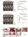
Figure 2.. S. aureus skin infection in vivo imaging and histology.
Three 8-mm in length, parallel scalpel wounds on the backs of (A-D) LysM-EGFP mice or (E) C57BL/6 mice inoculated with 2 × 10⁶ colony-forming units (CFUs) per 10 μl of Staphylococcus aureus or no bacteria (none). (A) Representative photographs of in vivo S. aureus bioluminescence. (B) In vivo S. aureus burden as measured by in vivo bioluminescence imaging (mean total flux (photons per second) ± SEM) (logarithmic scale). (C) Representative photographs of in vivo EGFP-neutrophil fluorescence. (D) In vivo fluorescence imaging of EGFP-neutrophil infiltration (mean total flux (photons per second) ± SEM). (E) Representative photomicrographs of sections from skin punch biopsies at 1 day after wounding ± S. aureus infection labeled with hematoxylin and eosin (H&E) stain, anti-Gr-1 mAb (neutrophil marker), and Gram stain. Scale bars = 150 μm. This figure was derived from (Cho et al., 2011).

Similar articles

Cited by

References

    1. Al-Nuaimi Y, Baier G, Watson RE, Chuong CM, Paus R. The cycling hair follicle as an ideal systems biology research model. Exp Dermatol 2010;19(8):707–13. - PMC - PubMed
    1. Alexander H, Brown S, Danby S, Flohr C. Research Techniques Made Simple: Transepidermal Water Loss Measurement as a Research Tool. J Invest Dermatol 2018;138(11):2295–300.e1. - PubMed
    1. Anderson LS, Reynolds MB, Rivara KR, Miller LS, Simon SI. A Mouse Model to Assess Innate Immune Response to Staphylococcus aureus Infection. J Vis Exp 2019(144). - PMC - PubMed
    1. Archer NK, Wang Y, Ortines RV, Liu H, Nolan SJ, Liu Q, et al. Preclinical Models and Methodologies for Monitoring Staphylococcus aureus Infections Using Noninvasive Optical Imaging. Methods Mol Biol 2020;2069:197–228. - PMC - PubMed
    1. Asai A, Tsuda Y, Kobayashi M, Hanafusa T, Herndon DN, Suzuki F. Pathogenic role of macrophages in intradermal infection of methicillin-resistant Staphylococcus aureus in thermally injured mice. Infect Immun 2010;78(10):4311–9. - PMC - PubMed

Publication types