Review
. 2021 Apr:110:83-97.
doi: 10.1016/j.humpath.2020.04.012. Epub 2020 May 11.

Data set for the reporting of pheochromocytoma and paraganglioma: explanations and recommendations of the guidelines from the International Collaboration on Cancer Reporting

Affiliations
Review

Data set for the reporting of pheochromocytoma and paraganglioma: explanations and recommendations of the guidelines from the International Collaboration on Cancer Reporting

Lester D R Thompson et al. Hum Pathol. 2021 Apr.

Abstract

Background and objectives: The International Collaboration on Cancer Reporting (ICCR) is a not-for-profit to develop evidence-based, internationally agreed-upon standardized data sets for each anatomic site, to be used throughout the world. Providing global standardization of pathology tumor classification, staging, and other reporting elements will lead to improved patient management and enhanced epidemiological research.

Methods: Pheochromocytoma and paraganglioma are uncommon and are frequently overlooked in registry data sets. Malignant criteria have previously been defined only when there was metastatic disease.

Results: With recent recognition of a significant inheritance association and the development of risk stratification tools, this data set was created in order to obtain more meaningful outcomes and management data, using similar criteria across the global pathology community. Issues related to key core and non-core elements, especially clinical hormonal status, familial history, tumor focality, proliferative fraction, adverse or risk stratification features, and ancillary techniques, are discussed in the context of daily application to these types of specimens.

Conclusions: The ICCR data set, developed by an international panel of endocrine organ specialists, establishes a pathology-standardized reporting guide for pheochromocytoma and paraganglioma.

Keywords: Checklist; Data set; ICCR; Paraganglioma; Pheochromocytoma; Structured report; Synoptic reporting.


Figures

Fig. 1
Fig. 1
A gross specimen of adrenal gland pheochromocytoma with a yellow, necrotic center. The rim of the residual adrenal cortex can be seen at the edge of the tumor.
Fig. 2
Fig. 2
A carotid body paraganglioma at the bifurcation of the carotid artery is highlighted by angiography (A), vascular magnetic resonance (MR) imaging after contrast administration (B), T1-weighted MR imaging (C), or MR imaging after contrast administration (D).
Fig. 3
Fig. 3
The biosynthetic pathway of catecholamines is demonstrated, including the enzymes necessary for synthesis. Metabolites are also shown as these are frequently used in testing. Not all pathways are illustrated, and not all intermediate steps are included. Tyrosinase is not expressed in the adrenal medulla or paraganglia, but is shown to illustrate the parallel utilization of tyrosine to produce melanin in melanocytes. Red circles show the structural region altered by the preceding enzyme’s action.
Fig. 4
Fig. 4
A, A coronal computed tomography image demonstrating bilateral carotid body paragangliomas in a syndrome-associated patient. B, A neck paraganglioma containing embolic material with associated foreign body giant cell reaction.
Fig. 5
Fig. 5
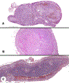
A, Multifocal tumors showing two topographically separate pheochromocytomas in the same adrenal gland. B, A microscopic pheochromocytoma identified in a background of diffuse hyperplasia. C, Diffuse adrenal medullary hyperplasia, with the medullary zone much greater than one-third of the gland thickness.
Fig. 6
Fig. 6
A, The overall size of this pheochromocytoma is large as a consequence of hemorrhage and necrosis. Fat has been removed to aid in accurate measuring. B, This is a gross photograph of a composite tumor (yellow) and medullary hyperplasia (brown), with measurements from each separate component documented.
Fig. 7
Fig. 7
A, The classical appearance of a paraganglioma with well-developed zellballen architecture. B, A composite ganglioneuroma and pheochromocytoma.
Fig. 8
Fig. 8
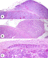
A, The pheochromocytoma invades through the capsule of the adrenal and expands into the adjacent adipose tissue. B, There is a well-developed capsule with tumor in the adjacent adipose tissue. C, Tumor cells are noted within an oval lymphatic endothelium-lined space in the capsule of a paraganglioma.
Fig. 9
Fig. 9
A, A Ki-67 proliferation index demonstrating >3% of the neoplastic nuclei are reactive. B, Histologic confirmation of lymph node metastasis from a paraganglioma.
Fig. 10
Fig. 10
The top row shows increasing cellularity, from low cellularity (A) to intermediate cellularity (B) to high cellularity (C) taken at the same magnification. The lower row demonstrates large nest size in comparison with usual smaller nests of paraganglioma (D) and tumor comedonecrosis (E).
Fig. 11
Fig. 11
Various ancillary studies aid in the diagnosis and evaluation of paraganglioma and pheochromocytoma. A, Strong, diffuse, nuclear INSM1. B, Strong, diffuse, nuclear reaction with GATA3. C, Tyrosine hydroxylase in a cytoplasmic distribution. D, Dopamine beta-hydroxylase with a cytoplasmic distribution. E, Sustentacular supporting S100 proteinepositive cells. F, Loss of SDHB in the neoplastic cells, with a strong internal control. SDHB, succinate dehydrogenase B.

References

    1. Srigley J, Lankshear S, Brierley J, McGowan T, Divaris D, Yurcan M, et al. Closing the quality loop: facilitating improvement in oncology practice through timely access to clinical performance indicators. J Oncol Pract 2013;9:e255–61. - PubMed
    1. Ellis DW, Srigley J. Does standardised structured reporting contribute to quality in diagnostic pathology? The importance of evidence-based datasets. Virchows Arch 2016;468:51–9. - PubMed
    1. Tischler AS, Asa SL, Clifton-Bligh R, de Krijger RR, Kimura N, Komminoth P, et al. Phaeochromocytoma and Paraganglioma Histopathology Reporting Guide. Sydney, Australia: International Collaboration on Cancer Reporting; 2019.
    1. Merlin T, Weston A, Tooher R. Extending an evidence hierarchy to include topics other than treatment: revising the Australian ‘levels of evidence. BMC Med Res Methodol 2009;9:34. - PMC - PubMed
    1. Giordano T, Berney D, de Krijger R, Erickson LA, Fassnacht M, Mete O, et al. Carcinoma of the Adrenal Cortex Histopathology Reporting Guide. Sydney, Australia: International Collaboration on Cancer Reporting; 2019.

Publication types

MeSH terms