Review
. 2020 Jun 5:20:739-753.
doi: 10.1016/j.omtn.2020.04.012. Epub 2020 Apr 29.

Mutation-Directed Therapeutics for Neurofibromatosis Type I

Affiliations
Review

Mutation-Directed Therapeutics for Neurofibromatosis Type I

Andre Leier et al. Mol Ther Nucleic Acids. .

Abstract

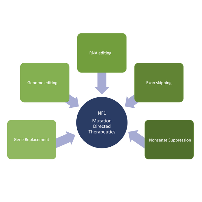
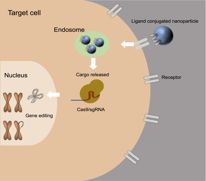
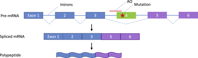
Significant advances in biotechnology have led to the development of a number of different mutation-directed therapies. Some of these techniques have matured to a level that has allowed testing in clinical trials, but few have made it to approval by drug-regulatory bodies for the treatment of specific diseases. While there are still various hurdles to be overcome, recent success stories have proven the potential power of mutation-directed therapies and have fueled the hope of finding therapeutics for other genetic disorders. In this review, we summarize the state-of-the-art of various therapeutic approaches and assess their applicability to the genetic disorder neurofibromatosis type I (NF1). NF1 is caused by the loss of function of neurofibromin, a tumor suppressor and downregulator of the Ras signaling pathway. The condition is characterized by a variety of phenotypes and includes symptoms such as skin spots, nervous system tumors, skeletal dysplasia, and others. Hence, depending on the patient, therapeutics may need to target different tissues and cell types. While we also discuss the delivery of therapeutics, in particular via viral vectors and nanoparticles, our main focus is on therapeutic techniques that reconstitute functional neurofibromin, most notably cDNA replacement, CRISPR-based DNA repair, RNA repair, antisense oligonucleotide therapeutics including exon skipping, and nonsense suppression.


Figures

None
Graphical abstract
Figure 1
Figure 1
Nanoparticle Delivery Delivery of CRISPR-Cas9 gene editing to a target cell through ligand-conjugated nanoparticles is shown. Nanoparticles recognize targeted cells through ligand-receptor interaction and penetrate into cells through endocytosis. After escaping the endosome, nanoparticles release cargo CRISPR-Cas9, which, in turn, translocates into the nucleus and exerts its gene editing function.
Figure 2
Figure 2
Trans-Splicing Ribozymes (A) Secondary structure of the group I intron ribozyme from Tetrahymena thermophila, in its re-designed format for 3′-replacement trans-splicing. All splice sites are indicated by black triangles. (A) The ribozyme (black) recognizes the target site on the substrate (red) with its 5′-terminus and a 5′-terminal extension (green) that enhances trans-splicing efficiency. During the trans-splicing reaction (gray arrow) the ribozyme 3′-tail (blue) replaces the substrate 3′-fragment. This reaction can be used to repair genetic mutations on the RNA level if the mutation is located in the substrate 3′-fragment and the ribozyme’s 3′-tail contains the correct sequence. (B–D) Three different modes of trans-splicing ribozymes. All splice sites are indicated by black triangles. (B) 3′-Replacement ribozymes (black) bind to the target site on the substrate (red) with their 5′-terminus. During the trans-splicing reaction their 3′-tail (blue) replaces the 3′-fragment of the substrate. (C) 5′-Replacement ribozymes (black) bind to the target site on the substrate (red) with their 3′-terminus. Their 5′-tail replaces the 5′-fragment of the substrate. (D) Internal fragment deletion ribozymes (black) bind to two target sites on the substrate (red/blue/red) with their 5′-terminus and with their 3′-terminus. Trans-splicing removes the fragment between the splice sites (blue) and joins the flanking portions of the product (red).
Figure 3
Figure 3
Exon Skipping Exon skipping is a form of RNA splicing where faulty, misaligned, or targeted coding sections of genetic sequence (exons) are “skipped,” leading to truncated but functional proteins. Skipping is mediated by antisense oligonucleotides (AOs) (denoted in red), which are short, synthetic pieces of modified RNA or DNA that hybridize to RNA via base pairing, causing evasion of the splicing machinery, i.e., skipping. Here, the top line denotes the exon-intron structure of a gene with a mutation in exon 4 (denoted with a red star). When an AO to exon 4 hybridizes, it masks the exon 4 splice site so that the splicing machinery does not recognize it and a transcript is created without it (middle line). This results in a protein that is missing exon 4 (bottom line). If this protein maintains the reading frame and exon 4 does not contain a domain critical for protein function, the product may retain function.
Figure 4
Figure 4
Nonsense Suppression Therapeutics (A) When normal translation occurs, a full-length functional polypeptide is generated. Translation is terminated when the ribosome encounters a normal stop codon (UAA is denoted in red). (B) When a ribosome encounters a premature termination codon (PTC) that was generated by a nonsense mutation, the ribosome terminates the transcript and a truncated protein is produced. Generally, the truncated transcript undergoes NMD, which lowers the cellular levels of this protein product. (C) Aminoglycosides and other small molecules can induce the ribosome to readthrough a PTC. If this occurs, one of three possible products may be made; these are not mutually exclusive. The correct amino acid may be inserted in place of the PTC; this leads to a full-length functional protein. Alternatively, a different amino acid may be inserted in place of the PTC; this leads to a missense mutation. This missense will either lead to a functional protein or a non-functional protein.

Similar articles

Cited by

References

    1. Friedman J.M., Gutmann D.H., MacCollin M., Riccardi V.M., editors. Neurofibromatosis: Phenotype, Natural History, and Pathogenesis. Third Edition. Johns Hopkins University Press; 1999.
    1. Kallionpää R.A., Uusitalo E., Leppävirta J., Pöyhönen M., Peltonen S., Peltonen J. Prevalence of neurofibromatosis type 1 in the Finnish population. Genet. Med. 2018;20:1082–1086. - PubMed
    1. Buratti E., Baralle D. Exon skipping mutations in neurofibromatosis. Methods Mol. Biol. 2012;867:65–76. - PubMed
    1. Vandenbroucke I., Callens T., De Paepe A., Messiaen L. Complex splicing pattern generates great diversity in human NF1 transcripts. BMC Genomics. 2002;3:13. - PMC - PubMed
    1. Thomson S.A., Wallace M.R. RT-PCR splicing analysis of the NF1 open reading frame. Hum. Genet. 2002;110:495–502. - PubMed