

Retinopathy Phenotypes in Type 2 Diabetes with Different Risks for Macular Edema and Proliferative Retinopathy
- PMID: 32408522
- PMCID: PMC7290313
- DOI: 10.3390/jcm9051433
Retinopathy Phenotypes in Type 2 Diabetes with Different Risks for Macular Edema and Proliferative Retinopathy
Abstract
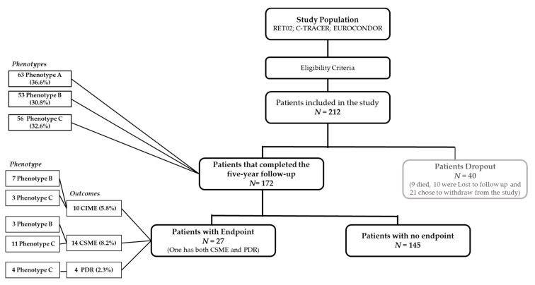
Our group reported that three diabetic retinopathy (DR) phenotypes: A, characterized by low microaneurysm turnover (MAT < 6) and normal central retinal thickness (CRT); B, low MAT (<6) and increased CRT, and C, high MAT (≥6), present different risks for development of macular edema (DME) and proliferative retinopathy (PDR). To test these findings, 212 persons with type 2 diabetes (T2D) and mild nonproliferative retinopathy (NPDR), one eye per person, were followed for five years with annual visits. Of these, 172 completed the follow-up or developed an outcome: PDR or DME (considering both clinically significant macular edema (CSME) and center-involved macular edema (CIME)). Twenty-seven eyes (16%) developed either CSME (14), CIME (10), or PDR (4), with one eye developing both CSME and PDR. Phenotype A showed no association with development of vision-threatening complications. Seven eyes with phenotype B and three with phenotype C developed CIME. Phenotype C showed higher risk for CSME development, with 17.41 odds ratio (p = 0.010), compared with phenotypes A + B. All eyes that developed PDR were classified as phenotype C. Levels of HbA1c and triglycerides were increased in phenotype C (p < 0.001 and p = 0.018, respectively). In conclusion, phenotype C identifies eyes at higher risk for development of CSME and PDR, whereas phenotype A identifies eyes at very low risk for vision-threatening complications.
Keywords: diabetes; macular edema; proliferative retinopathy; retinopathy.
Conflict of interest statement
I.P.M., M.H.M., A.L.M., T.S. and A.C.–V.M. do not have financial disclosures. J.F.: member of Advisory board for Alimera, Allergan, Bayer, Bhoeringer, and Novartis; J.C.–V. reports grants from Carl Zeiss Meditec outside the submitted work and is consultant for Alimera Sciences, Allergan, Bayer, Gene Signal, Novartis, Pfizer, Precision Ocular Ltd., Roche, Sanofi–Aventis, Vifor Pharma, and Carl Zeiss Meditec.
Figures

References
-
- Lobo C.L., Bernardes R.C., Figueira J.P., Faria De Abreu J.R., Cunha–Vaz J.G. Three–Year Follow–up Study of Blood–Retinal Barrier and Retinal Thickness Alterations in Patients with Type 2 Diabetes Mellitus and Mild Nonproliferative Diabetic Retinopathy. Arch. Ophthalmol. 2004;122:211–217. doi: 10.1001/archopht.122.2.211. - DOI - PubMed
Grants and funding
LinkOut - more resources
Full Text Sources
Research Materials
Miscellaneous
