. 2020 May 12;9(5):1201.
doi: 10.3390/cells9051201.

Progerin Expression Induces Inflammation, Oxidative Stress and Senescence in Human Coronary Endothelial Cells

Affiliations

Progerin Expression Induces Inflammation, Oxidative Stress and Senescence in Human Coronary Endothelial Cells

Guillaume Bidault et al. Cells. .

Abstract

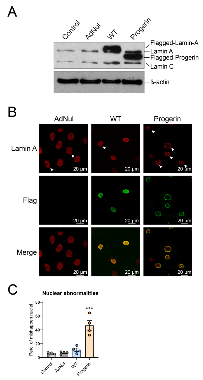
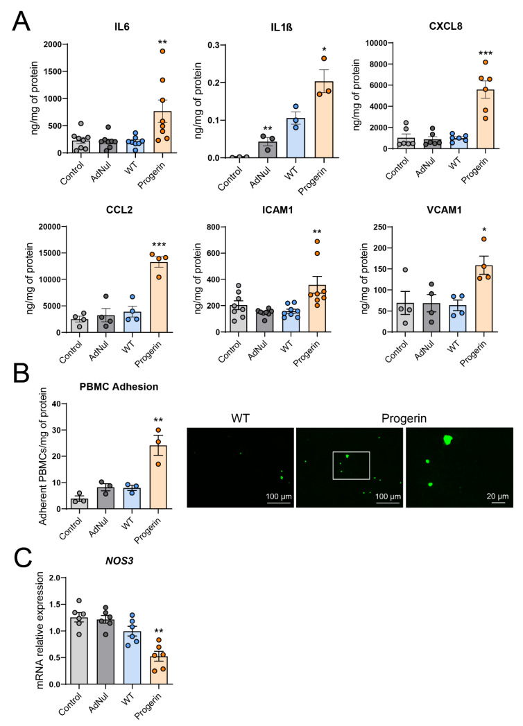
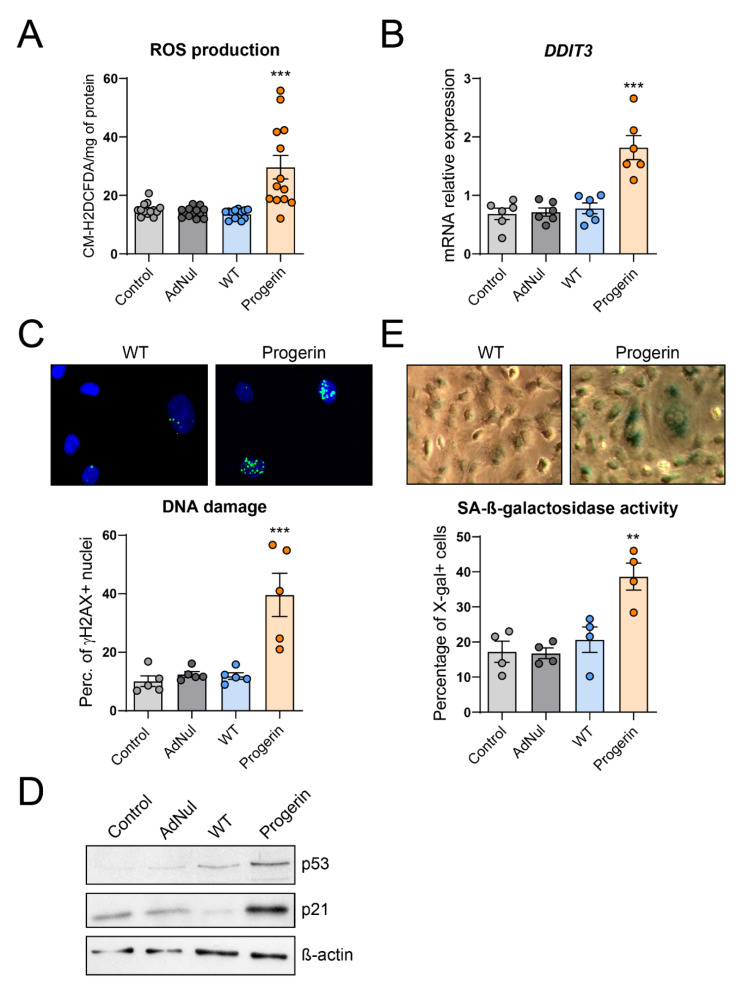
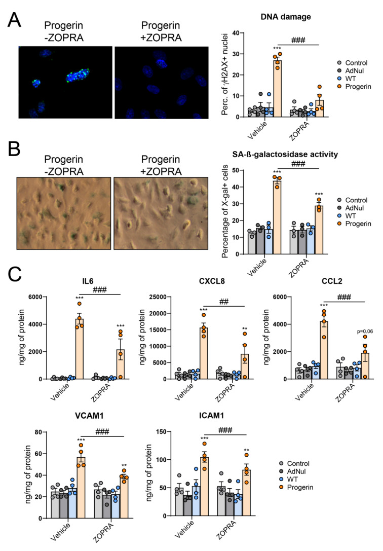
Hutchinson-Gilford progeria syndrome (HGPS) is a rare premature aging disorder notably characterized by precocious and deadly atherosclerosis. Almost 90% of HGPS patients carry a LMNA p.G608G splice variant that leads to the expression of a permanently farnesylated abnormal form of prelamin-A, referred to as progerin. Endothelial dysfunction is a key determinant of atherosclerosis, notably during aging. Previous studies have shown that progerin accumulates in HGPS patients' endothelial cells but also during vascular physiological aging. However, whether progerin expression in human endothelial cells can recapitulate features of endothelial dysfunction is currently unknown. Herein, we evaluated the direct impact of exogenously expressed progerin and wild-type lamin-A on human endothelial cell function and senescence. Our data demonstrate that progerin, but not wild-type lamin-A, overexpression induces endothelial cell dysfunction, characterized by increased inflammation and oxidative stress together with persistent DNA damage, increased cell cycle arrest protein expression and cellular senescence. Inhibition of progerin prenylation using a pravastatin-zoledronate combination partly prevents these defects. Our data suggest a direct proatherogenic role of progerin in human endothelial cells, which could contribute to HGPS-associated early atherosclerosis and also potentially be involved in physiological endothelial aging participating to age-related cardiometabolic diseases.

Keywords: Hutchinson–Gilford progeria syndrome; LMNA; aging; atherosclerosis; endothelial dysfunction; inflammation; lamin A; prenylation; progerin.


Conflict of interest statement

The authors declare no conflict of interest.

Figures

Figure 1
Figure 1
Expression and localization of exogenous lamin A and progerin. Early-confluent human coronary artery endothelial cells (HCAECs) were transduced or not with Flag-tagged recombinant adenovirus containing WT-prelamin A or progerin for 72 h, or with an empty vector (AdNul). (A) Western blot analysis of lamin A and C and progerin. β-actin was used as a loading control. Representative picture of n = 4 experiments. (B) Representative pictures of transduced HCAECs stained with Flag (green) and lamin-A (red) antibodies. Cells were observed by confocal microscopy at 100× magnification. Examples of misshapen nuclei are indicated with a white arrow. (C) Quantification of misshapen nuclei as percentage of total nuclei. Data are expressed as the mean ± standard error of mean (SEM) and statistical difference is determined using analysis of variance (ANOVA) followed by a Dunnett post hoc test. *** p < 0.001 vs. WT.
Figure 2
Figure 2
Progerin induces endothelial cells inflammation and dysfunction. Early-confluent HCAECs were transduced or not with Flag-tagged recombinant adenovirus containing WT-prelamin A or progerin for 72 h or with an empty vector. (A) Twenty-four hour secretion of the proinflammatory cytokines IL6, IL1β, of the chemokines CXCL8 and CCL2 and of the adhesion molecules ICAM1 and VCAM1. (B) Adhesion assay of peripheral blood mononuclear cells (PBMCs) from healthy donors on transduced and control endothelial cells was quantified as the number of adherent PBMC/mg of protein (left panel). Representative pictures are shown in the right panel with a magnification of the white squared area (C) Relative mRNA expression of the endothelial nitric oxide synthase gene (NOS3). Data are expressed as the mean ± SEM and statistical difference is determined using ANOVA followed by a Dunnett post hoc test. * p < 0.05, ** p < 0.01, *** p < 0.001 vs. WT.
Figure 3
Figure 3
Endothelial progerin expression induces oxidative stress, DNA damage and cellular senescence. Early-confluent HCAECs were transduced or not with Flag-tagged recombinant adenovirus containing WT-prelamin A or progerin or with an empty vector. (A) Reactive oxygen species (ROS) production was assessed by the oxidation of 5-6-chloromethyl-2,7-dichlorodihydro-fluorescein diacetate (CM-H2DCFDA). (B) Relative mRNA expression of DDIT3. (C) DNA double-strand breaks (DSBs) were studied by staining HCAECs with Ser139-phosphorylated histone variant H2A (γ-H2AX, in green) and di-amidino-2-phenylindole hydrochloride (DAPI) (in blue) (upper panel) and evaluated as the percentage of γ-H2AX–positive cells (40–200 cells per experiment) (lower panel). (D) Protein expression of the cell cycle arrest proteins p53 and p21. Representative picture of n = 3 experiments. (E) Senescence-associated (SA)-β-galactosidase activity was assessed by the percentage of 5-bromo-4-chloro-3-indolyl-β-d-galactopyranoside (X-gal)-stained HCAECs (in blue) at pH6. Representative micrographs are shown (upper panel). Data are expressed as the mean ± SEM and statistical difference determined using ANOVA followed by a Dunnett post hoc test. ** p < 0.01, *** p < 0.001 vs. WT.
Figure 4
Figure 4
Inhibition of progerin prenylation partially prevents endothelial cells senescence, inflammation and secretion of adhesion molecules. Early-confluent HCAECs were transduced or not with Flag-tagged recombinant adenovirus containing WT-prelamin A or progerin for 72 h and immediately treated with zoledronate and pravastatin (ZOPRA). (A) DNA DSBs were studied by staining HCAECs with γ-H2AX (in green) and DAPI (in blue). Representative micrographs of HCAECs expressing progerin are shown (left panel). DNA DSBs were quantified as the percentage of γ-H2AX–positive cells (40–200 cells per experiment) (right panel). (B) Senescence-associated (SA)-β-galactosidase activity was assessed as the percentage of X-gal–stained cells (blue) at pH6 (right panel). Representative micrographs are shown (left panel). (C) 24 h-secretion of the proinflammatory cytokine IL6, of the chemokines CXCL8 and CCL2 and of the adhesion molecules ICAM1 and VCAM1. Data are expressed as the mean ± SEM and statistical difference was determined using two-way ANOVA followed by a Tukey post hoc test to assess differences against WT-prelamin A overexpression or a Sidak post hoc test to determine ZOPRA effect for each condition. *** p < 0.001 vs. WT. ## p < 0.01, ### p < 0.001 vs. vehicle-treated cells.

References

    1. De Sandre-Giovannoli A., Bernard R., Cau P., Navarro C., Amiel J., Boccaccio I., Lyonnet S., Stewart C.L., Munnich A., Le Merrer M., et al. Lamin a truncation in Hutchinson–Gilford progeria. Science. 2003;300:2055. doi: 10.1126/science.1084125. - DOI - PubMed
    1. Eriksson M., Brown W.T., Gordon L.B., Glynn M.W., Singer J., Scott L., Erdos M.R., Robbins C.M., Moses T.Y., Berglund P., et al. Recurrent de novo point mutations in lamin A cause Hutchinson–Gilford progeria syndrome. Nature. 2003;423:293–298. doi: 10.1038/nature01629. - DOI - PMC - PubMed
    1. Merideth M.A., Gordon L.B., Clauss S., Sachdev V., Smith A.C., Perry M.B., Brewer C.C., Zalewski C., Kim H.J., Solomon B., et al. Phenotype and course of Hutchinson–Gilford progeria syndrome. N. Engl. J. Med. 2008;358:592–604. doi: 10.1056/NEJMoa0706898. - DOI - PMC - PubMed
    1. Dechat T., Adam S.A., Taimen P., Shimi T., Goldman R.D. Nuclear lamins. Cold Spring Harb. Perspect. Biol. 2010;2:a000547. doi: 10.1101/cshperspect.a000547. - DOI - PMC - PubMed
    1. Corrigan D.P., Kuszczak D., Rusinol A.E., Thewke D.P., Hrycyna C.A., Michaelis S., Sinensky M.S. Prelamin A endoproteolytic processing in vitro by recombinant Zmpste24. Biochem. J. 2005;387:129–138. doi: 10.1042/BJ20041359. - DOI - PMC - PubMed

Publication types

LinkOut - more resources