Case Reports
. 2020 Dec;26(6):821-825.
doi: 10.1177/1591019920926333. Epub 2020 May 14.

Endovascular coiling of large mastoid emissary vein causing pulsatile tinnitus

Affiliations
Case Reports

Endovascular coiling of large mastoid emissary vein causing pulsatile tinnitus

Mohamad Abdalkader et al. Interv Neuroradiol. 2020 Dec.

Abstract

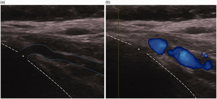
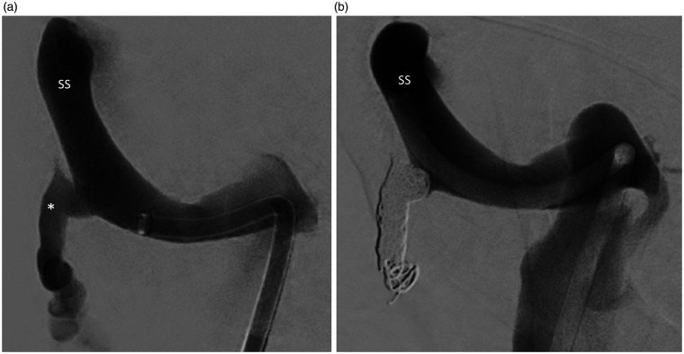
The association of large mastoid emissary veins and pulsatile tinnitus has been reported. However, therapeutic options for this condition remain limited. We report a case of endovascular coiling of a large mastoid emissary vein in a patient with disabling pulsatile tinnitus with significant improvement of symptoms. To our knowledge, endovascular coiling of large mastoid emissary vein causing pulsatile tinnitus has not been reported.

Keywords: Pulsatile tinnitus; coil embolization; mastoid emissary vein.


Conflict of interest statement

Declaration of conflicting interests: The author(s) declared no potential conflicts of interest with respect to the research, authorship, and/or publication of this article.

Figures

Figure 1.
Figure 1.
Axial nonenhanced CT scan at the level of the posterior fossa showing a large right mastoid emissary canal measuring up to 5 mm in its largest diameter. Note also of a prominent left mastoid emissary vein of approximately 2 mm.
Figure 2.
Figure 2.
Ultrasound imaging in B-mode (a) and color Doppler (b) of the right mastoid area showing a right mastoid emissary vein exiting the right mastoid bone (dashed line) via the mastoid foramen (asterisk).
Figure 3.
Figure 3.
Venogram phase (right anterior oblique: 24°, caudal: 7°) of a right internal carotid artery injection before (a) and after (b) ultrasound probe compression over the right mastoid area showing obliteration of the right mastoid emissary vein (asterisk) after compression. Images are mildly degraded by motion artifacts. MB, mastoid bone; SS, sigmoid sinus; PAV, posterior auricular vein; IJV, internal jugular vein; SSS, superior sagittal sinus.
Figure 4.
Figure 4.
Venogram of the right sigmoid sinus (SS) (right anterior oblique: 25°, caudal: 16°) before (a) and after transvenous coiling (b) showing a dilated mastoid emissary vein (asterisk) connecting the right sigmoid sinus with the posterior auricular and occipital veins/suboccipital venous plexus with complete obliteration post coiling.

References

    1. Erlandsson SI and, Hallberg LR-M. Prediction of quality of life in patients with tinnitus. Br J Audiol 2000; 34: 11–19. - PubMed
    1. Zhao P, et al. CT evaluation of sigmoid plate dehiscence causing pulsatile tinnitus. Eur Radiol 2016; 26: 9–14. - PubMed
    1. Krishnan A, Mattox DE, Fountain AJ, et al. CT arteriography and venography in pulsatile tinnitus: preliminary results. AJNR Am J Neuroradiol 2006; 27: 1635–1638. - PMC - PubMed
    1. Braun JP, Tournade A. Venous drainage in the craniocervical region. Neuroradiology 1977; 13: 155–158. - PubMed
    1. Irmak MK, Korkmaz A, Erogul O. Selective brain cooling seems to be a mechanism leading to human craniofacial diversity observed in different geographical regions. Med Hypotheses 2004; 63: 974–979. - PubMed

Publication types