. 2020 May 14;17(1):156.
doi: 10.1186/s12974-020-01814-4.

Anti-IL-20 antibody improved motor function and reduced glial scar formation after traumatic spinal cord injury in rats

Affiliations

Anti-IL-20 antibody improved motor function and reduced glial scar formation after traumatic spinal cord injury in rats

Jung-Shun Lee et al. J Neuroinflammation. .

Abstract

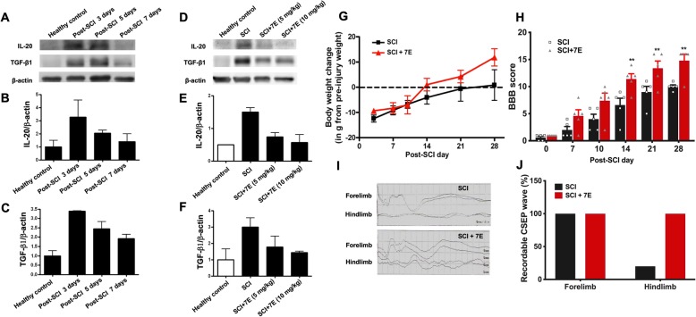
Background: Spinal cord injury (SCI) causes devastating neurological consequences, which can result in partial or total paralysis. Irreversible neurological deficits and glial scar formation are characteristic of SCI. Inflammatory responses are a major component of secondary injury and play a central role in regulating the pathogenesis of SCI. IL-20 is a proinflammatory cytokine involved in renal fibrosis and liver cirrhosis through its role in upregulating TGF-β1 production. However, the role of IL-20 in SCI remains unclear. We hypothesize that IL-20 is upregulated after SCI and is involved in regulating the neuroinflammatory response.

Methods: The expression of IL-20 and its receptors was examined in SCI rats. The regulatory roles of IL-20 in astrocytes and neuron cells were examined. The therapeutic effects of anti-IL-20 monoclonal antibody (mAb) 7E in SCI rats were evaluated.

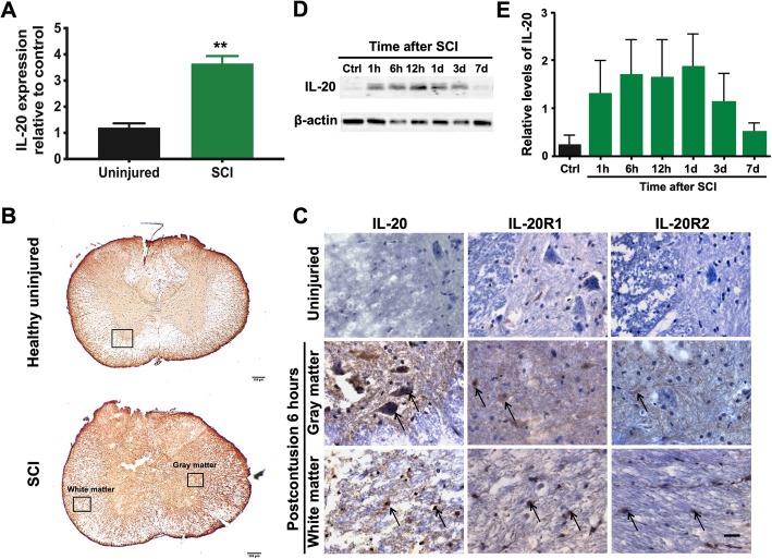
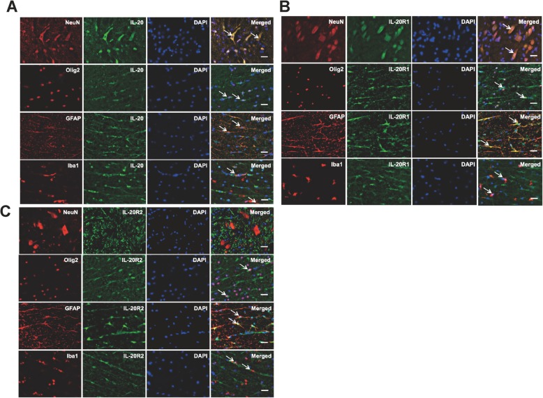
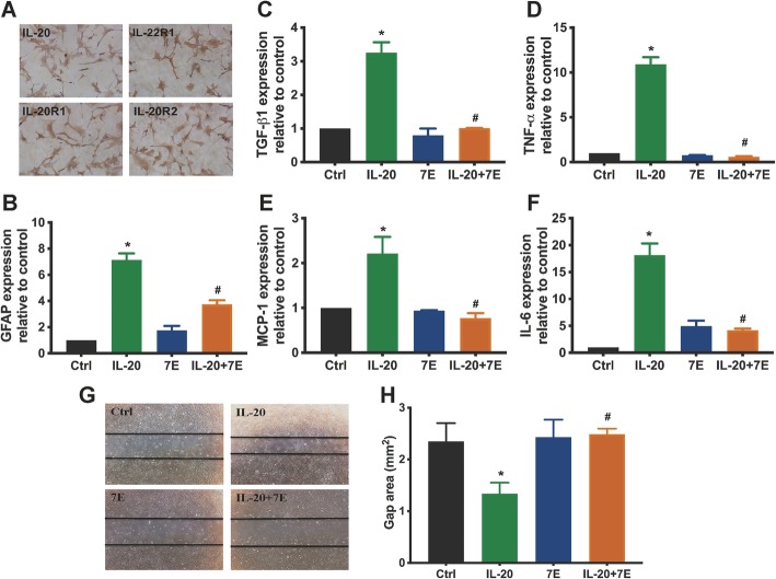
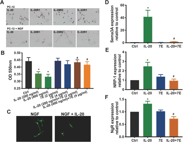
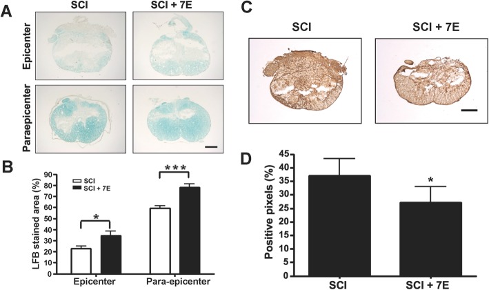
Results: Immunofluorescence staining showed that IL-20 and its receptors were expressed in astrocytes, oligodendrocytes, and microglia in the spinal cord after SCI in rats. In vitro, IL-20 enhanced astrocyte reactivation and cell migration in human astrocyte (HA) cells by upregulating glial fibrillary acidic protein (GFAP), TGF-β1, TNF-α, MCP-1, and IL-6 expression. IL-20 inhibited cell proliferation and nerve growth factor (NGF)-derived neurite outgrowth in PC-12 cells through Sema3A/NRP-1 upregulation. In vivo, treating SCI rats with anti-IL-20 mAb 7E remarkably inhibited the inflammatory responses. 7E treatment not only improved motor and sensory functions but also improved spinal cord tissue preservation and reduced glial scar formation in SCI rats.

Conclusions: IL-20 might regulate astrocyte reactivation and axonal regeneration and result in the secondary injury in SCI. These findings demonstrated that IL-20 may be a promising target for SCI treatment.

Keywords: IL-20; Neuroinflammation; SCI.


Conflict of interest statement

The authors declare no competing interests.

Figures

Fig. 1
Fig. 1
Upregulation of IL-20 after spinal cord injury (SCI). a Spinal cord tissues from healthy rats (uninjured; N = 4) and SCI rats (N = 6) were collected at 3 days post-SCI. Total RNA was isolated and the transcripts of IL-20 were measured using RT-qPCR with specific primers. GAPDH was an internal control. **P < 0.01 compared with the healthy uninjured controls. Data are expressed as mean ± SD. b Spinal cord sections obtained from healthy uninjured rats (N = 5) and SCI rats (N = 5) at 6 h after the initial injury. Scale bars = 200 μm. c Spinal cord tissue samples were stained with anti-IL-20 mAb using immunohistochemical staining. Staining for IL-20 was positive in the injured spinal cord, not only in the gray matter, but also in the white matter. Scale bars = 500 μm. d Spinal cord tissue from healthy control rats (N = 5) and SCI rats (N = 5; each time point) were collected at the indicated time points post-SCI. Tissue lysates were analyzed through immunoblotting with specific antibodies against IL-20. β-actin was an internal control. e Relative levels of IL-20 quantified by densitometric analysis using ImageJ software. Data are expressed as mean ± SD and are representative of three independent experiments
Fig. 2
Fig. 2
Expression of IL-20, IL-20R1, and IL-20R2 in spinal cord tissues after SCI. The transverse sections around the interface between gray and white matters of the spinal cord obtained from SCI rats (N = 5). a Double immunofluorescence staining of IL-20 (green) with markers for specific neural cell types (red) including NeuN (neurons), Olig2 (oligodendrocytes), GFAP (astrocytes), and Iba1 (microglia). Nuclei were counterstained with DAPI (blue). Co-localization of IL-20 with each cellular marker appears yellow in the merged image. Scale bars = 500 μm. b, c Double immunofluorescence staining of IL-20R1 (green) or IL-20R2 (green) with markers for specific neural cell types (red) as described above. Co-localization of IL-20R1 or IL-20R2 with each cellular marker appears yellow in the merged image. Scale bars = 500 μm. NeuN, neuronal nuclei; Olig2, oligodendrocyte transcription factor 2; GFAP, glial fibrillary acidic protein; Iba1, ionized calcium-binding adapter molecule 1. Data from one representative experiment of three independent experiments is shown
Fig. 3
Fig. 3
IL-20 promoted astrocyte activation in HA cell line. a Expression of IL-20 and its receptors (IL-20R1, IL-20R2, and IL-22R1) in HA cells was analyzed using immunocytochemistry staining. Data from one representative experiment of three independent experiments is shown. bf HA cells were treated with IL-20 (200 ng/ml), 7E (2 μg/ml), or IL-20 (200 ng/ml) plus 7E (2 μg/ml) for 4 h. Total RNA was isolated and the transcripts of GFAP, TGF-β1, TNF-α, MCP-1, and IL-6 were analyzed using RT-qPCR with specific primers. GAPDH was an internal control. *P < 0.05 compared with the untreated controls (Ctrl). #P < 0.05 compared with the IL-20-treated group. Data are expressed as mean ± SD and are representative of three independent experiments performed in triplicate. g, h Cell migration was evaluated using a wound-healing assay for 24 h. Wound closure was quantified by measuring the area remaining to be immigrated using ImageJ software. *P < 0.05 compared with the untreated controls (Ctrl). #P < 0.05 compared with the IL-20-treated group. Data are expressed as mean ± SD and are representative of three independent experiments performed in triplicate
Fig. 4
Fig. 4
IL-20 inhibited axonal regeneration. a Expression of IL-20 and its receptors (IL-20R1, IL-20R2, and IL-22R1) in PC-12 cells and nerve growth factor (NGF)-differentiated PC-12 cells were analyzed using immunocytochemistry staining. Data from one representative experiment of three independent experiments is shown. b NGF-differentiated PC-12 cells were incubated with IL-20, 7E, or IL-20 plus 7E for 72 h. Cell viability was determined using the MTT assay. *P < 0.05 compared with the untreated controls (Ctrl). #P < 0.05 compared with the corresponding controls. Data are expressed as mean ± SD and are representative of three independent experiments performed in quadruplicate. c PC-12 cells were treated with NGF or with NGF plus IL-20 (200 ng/ml). Immunofluorescence staining was performed using anti-β-tubulin mAb (green) to mark neurite outgrowth. Data from one representative experiment of three independent experiments is shown. d NGF-differentiated PC-12 cells were treated with IL-20 (200 ng/ml), 7E (2 μg/ml), or IL-20 (200 ng/ml) plus 7E (2 μg/ml) for 6 h. Total RNA was isolated, and the transcripts of Sema3A, NRP-1, and NgR were analyzed using RT-qPCR with specific primers. GAPDH was an internal control. *P < 0.05 compared with the untreated controls (Ctrl). #P < 0.05 compared with IL-20-treated group. Data are expressed as mean ± SD and are representative of three independent experiments performed in quadruplicate
Fig. 5.
Fig. 5.
Anti-IL-20 mAb 7E improved motor function and sensory function in SCI rats. a Spinal cord tissues from healthy control rats (N = 4) and SCI rats were collected at 3 days, 5 days, and 7 days post-SCI (N = 4; each time point). Tissue lysates were analyzed through immunoblotting with specific antibodies against IL-20 and TGF-β1. β-actin was an internal control. b, c Relative levels of IL-20 and TGF-β1 quantified by densitometric analysis using ImageJ software. Data are expressed as mean ± SD and are representative of three independent experiments. d Spinal cord tissues from healthy control and SCI rats treated with anti-IL-20 mAb 7E (5–10 mg/kg) were analyzed at 3 days post-SCI through immunoblotting with specific antibodies against IL-20 and TGF-β1 (N = 4/group). β-actin was an internal control. e, f Relative levels of IL-20 and TGF-β1 quantified by densitometric analysis using ImageJ software. Data are expressed as mean ± SD and are representative of three independent experiments. g SCI rats were treated with anti-IL-20 mAb 7E (10 mg/kg), and body weight was measured weekly (N = 5/group). h The Basso, Bresnahan, and Beattie (BBB) scoring method was applied to evaluate motor function in SCI control rats (N = 5/group) and SCI rats (N = 5/group) treated with anti-IL-20 mAb 7E (10 mg/kg) weekly post-SCI. **P < 0.01 compared with the SCI rats. Data are expressed as mean ± SD. Data from one representative experiment of three independent experiments is shown. i, j The cortical somatosensory-evoked potential (CSEP) waves were recorded to represent sensory function in SCI control rats and SCI rats treated with anti-IL-20 mAb 7E (10 mg/kg) at 28 days post-SCI (N = 5/group). Data from one representative experiment of three independent experiments is shown
Fig. 6
Fig. 6
Anti-IL-20 mAb 7E improved spinal cord tissue preservation and reduced glial scar formation. a Luxol fast blue (LFB) stain was used to identify myelinated tissues in SCI control rats and in SCI rats treated with anti-IL-20 mAb 7E (10 mg/kg) at 28 days post-SCI (N = 5/group). Scale bars = 500 μm. b Quantification of LFB stained areas. *P < 0.05, ***P < 0.001 compared with the SCI rats. Data are expressed as mean ± SD. Data from one representative experiment of three independent experiments is shown. c At 28 days post-SCI, spinal cord tissue samples from SCI control rats and from SCI rats treated with anti-IL-20 mAb 7E (10 mg/kg) were stained with anti-chondroitin sulfate proteoglycan (CSPG) mAb to evaluate glial scar formation (N = 5/group). Scale bars = 500 μm. d Quantification of CSPG-positive stained areas. *P < 0.05 compared with the SCI rats. Data are expressed as mean ± SD. Data from one representative experiment of three independent experiments is shown

References

    1. Orr MB, Gensel JC. Spinal cord injury scarring and inflammation: therapies targeting glial and inflammatory responses. Neurotherapeutics. 2018;15:541–553. doi: 10.1007/s13311-018-0631-6. - DOI - PMC - PubMed
    1. Kwon BK, Tetzlaff W, Grauer JN, Beiner J, Vaccaro AR. Pathophysiology and pharmacologic treatment of acute spinal cord injury. Spine J. 2004;4:451–464. doi: 10.1016/j.spinee.2003.07.007. - DOI - PubMed
    1. Ahuja CS, Wilson JR, Nori S, Kotter MRN, Druschel C, Curt A, Fehlings MG. Traumatic spinal cord injury. Nature Reviews Disease Primers. 2017;3:17018. doi: 10.1038/nrdp.2017.18. - DOI - PubMed
    1. Kwon B, Fisher CG, Dvorak M, Tetzlaff W. Strategies to Promote Neural Repair and Regeneration After Spinal Cord Injury. Spine (Phila Pa 1976) 2005;30(17 Suppl):S3–13. doi: 10.1097/01.brs.0000175186.17923.87. - DOI - PubMed
    1. Tator CH, Duncan EG, Edmonds VE, Lapczak LI, Andrews DF. Neurological recovery, mortality and length of stay after acute spinal cord injury associated with changes in management. Paraplegia. 1995;33:254–262. - PubMed