. 2020 Jul 10;295(28):9474-9489.
doi: 10.1074/jbc.RA120.013028. Epub 2020 May 14.

A specific oligosaccharide-binding site in the alternansucrase catalytic domain mediates alternan elongation

Affiliations

A specific oligosaccharide-binding site in the alternansucrase catalytic domain mediates alternan elongation

Manon Molina et al. J Biol Chem. .

Abstract

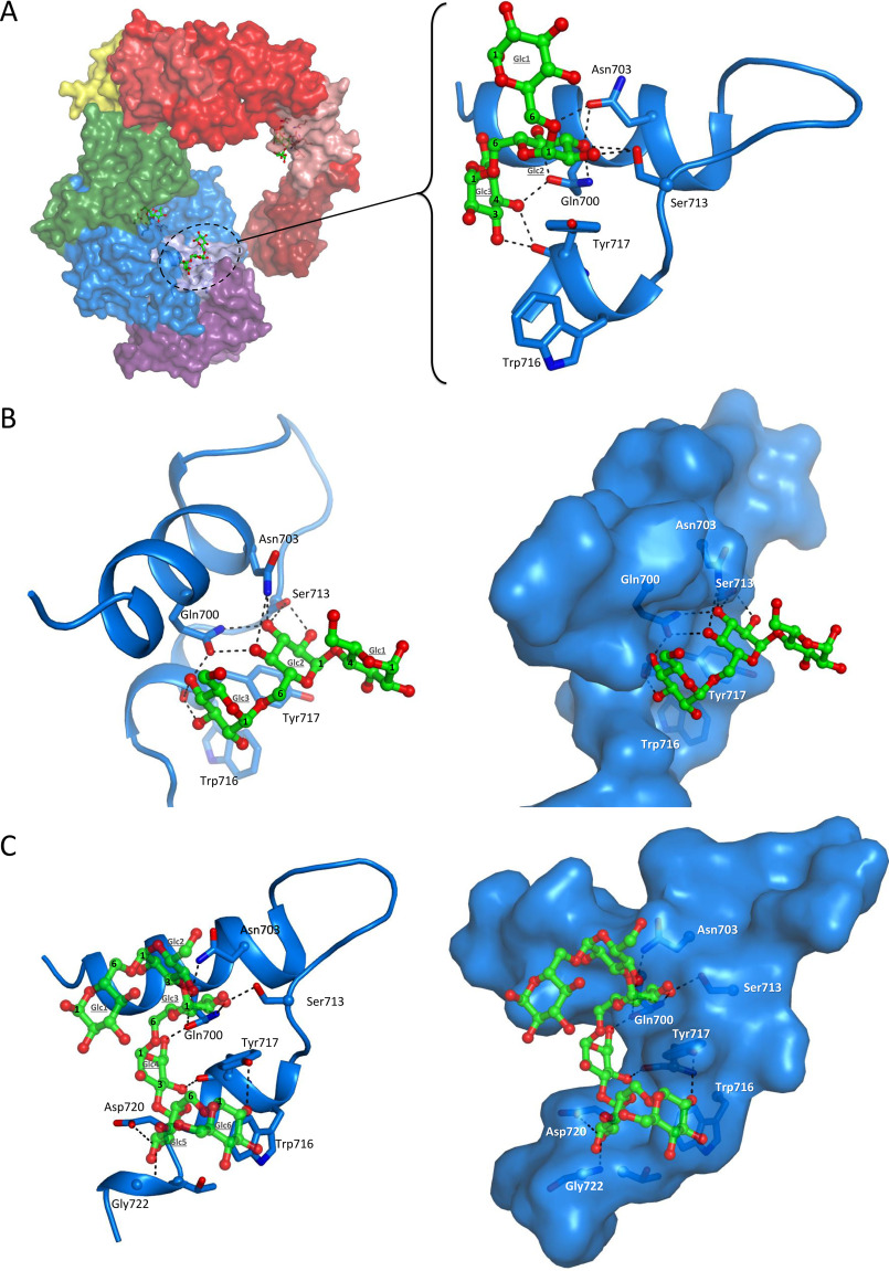
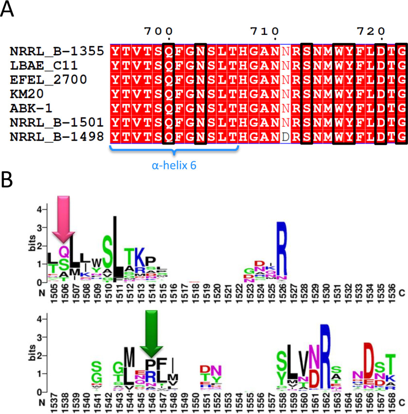
Microbial α-glucans produced by GH70 (glycoside hydrolase family 70) glucansucrases are gaining importance because of the mild conditions for their synthesis from sucrose, their biodegradability, and their current and anticipated applications that largely depend on their molar mass. Focusing on the alternansucrase (ASR) from Leuconostoc citreum NRRL B-1355, a well-known glucansucrase catalyzing the synthesis of both high- and low-molar-mass alternans, we searched for structural traits in ASR that could be involved in the control of alternan elongation. The resolution of five crystal structures of a truncated ASR version (ASRΔ2) in complex with different gluco-oligosaccharides pinpointed key residues in binding sites located in the A and V domains of ASR. Biochemical characterization of three single mutants and three double mutants targeting the sugar-binding pockets identified in domain V revealed an involvement of this domain in alternan binding and elongation. More strikingly, we found an oligosaccharide-binding site at the surface of domain A, distant from the catalytic site and not previously identified in other glucansucrases. We named this site surface-binding site (SBS) A1. Among the residues lining the SBS-A1 site, two (Gln700 and Tyr717) promoted alternan elongation. Their substitution to alanine decreased high-molar-mass alternan yield by a third, without significantly impacting enzyme stability or specificity. We propose that the SBS-A1 site is unique to alternansucrase and appears to be designed to bind alternating structures, acting as a mediator between the catalytic site and the sugar-binding pockets of domain V and contributing to a processive elongation of alternan chains.

Keywords: GH70; alternan; alternansucrase; biosourced; biotechnology; carbohydrate structure; dextran; enzyme catalysis; enzyme mechanism; glucansucrase; green chemistry; polymerase; polysaccharide; processivity; sugar-binding pockets; surface-binding site.


Conflict of interest statement

Conflict of interest—The authors declare that they have no conflicts of interest with the contents of this article.

Figures

Figure 1.
Figure 1.
Identification of the new sugar-binding sites: SBS-A1 and sugar-binding pocket V-B in ASR. The list of ligands found at these binding sites is indicated in the right panel with an illustration of the structure visible in the complexes. Gray, catalytic residues Asp635, Glu673, and Asp767. See Figs. S4 and S7 for electron density maps sugar-binding pocket V-B and in surface-binding site A1, respectively.
Figure 2.
Figure 2.
Complexes in sugar-binding pockets. A, crystal structure of isomaltotriose binding in pocket V-B (PDB code 6SYQ). B, possible location of isomaltotriose in pocket V-A as superimposed from pocket V-B. C, crystal structure of the OA bound in pocket V-B (PDB code 6T18). The probable interaction network is displayed as dashed lines. D, sequence alignment of the sugar-binding pockets identified in ASR, DSR-M, and DSR-E glucansucrases. Pink highlighted residues were shown to directly interact with sugar ligands in 3D structures. Residues in blue ovals have been mutated to Ala in this study.
Figure 3.
Figure 3.
HPSEC analysis of the mutants in the sugar-binding pockets of domain V. The reaction was from sucrose at 30 °C with 1 unit ml−1 of pure enzyme and 50 mm sodium acetate buffer, pH 5.75.
Figure 4.
Figure 4.
Affinity gel electrophoresis. A, affinity gel electrophoresis of ASRΔ2, ASRΔ5, and ASRΔ5-Y717A. DSR-MΔ1 and ΔN123-GBD-CD2 (branching sucrase) are used as a positive control (12, 14). 1% BSA and protein standard (ladder) are used as negative controls. The gels were made in the presence or absence of 0.45% (w/v) dextran 70,000 g mol−1 or alternan. B, affinity gel electrophoresis of ASRΔ2, ASRΔ2-Y158A, ASRΔ2-Y241A, ASRΔ2-Y158A/Y241A, and ASRΔ5. Protein standard (ladder) is used as negative control. The gels were made in the presence or absence of 0.45% (w/v) dextran 2,000,000 g mol−1 or 0.9% (w/v) alternan.
Figure 5.
Figure 5.
Complexes in SBS-A1. A, crystal structure of isomaltotriose binding in surface-binding site A1 (PDB code 6SYQ). Sucrose molecule in the active site was positioned from the superimposition of the ASR catalytic domain with GTF-180–sucrose complex (PDB code 3HZ3). B, crystal structure of panose binding in SBS-A1 (PDB code 6T16). C, crystal structure of OA binding in SBS-A1 (PDB code 6T18).
Figure 6.
Figure 6.
Reaction product analysis of SBS-A1 mutants. A, HPSEC chromatograms of the products synthesized by SBS-A1 mutants. B, HPAEC-PAD chromatogram of the oligoalternans produced from sucrose with ASRΔ2 and mutant Y717A. The reaction was from sucrose at 30 °C with 1 unit ml−1 of pure enzyme and 50 mm sodium acetate buffer, pH 5.75. C, HPAEC-PAD chromatogram of the acceptor reaction products from maltose, with ASRΔ2 and mutant Y717A. The reaction was from sucrose and maltose with sucrose:maltose mass ratio 2:1 at 30 °C with 1 unit ml−1 of pure enzyme and 50 mm sodium acetate buffer, pH 5.75. For detailed structures, see “Experimental procedures.”
Figure 7.
Figure 7.
Analysis of mutants combined to the Y717A mutation. A, HPSEC chromatograms of alternan populations produced with Y717A and the double mutants Y717A/Y158A mutant (pocket V-A) and Y717A/Y241A mutant (pocket V-B). The reaction was from sucrose at 30 °C with 1 unit ml−1 of pure enzyme and 50 mm sodium acetate buffer, pH 5.75. B, monitoring of polymer formation with time. The reaction was from sucrose at 30 °C with 1 unit ml−1 of pure enzyme and 50 mm sodium acetate buffer, pH 5.75. The production rate was calculated from 5 to 75 min (R2 of 0.997) and from 10 to 75 min (R2 of 0.999) for ASRΔ2 and ASRΔ2 Y717A, respectively.
Figure 8.
Figure 8.
SBS-A1 sequence comparison. A, alignment of the residues corresponding to SBS-A1 (in black boxes) in all characterized and putative alternansucrases. Only the strain name is indicated. The species was L. citreum or L. mensenteroides. Alignment created with ENDscript 2 (54). B, WebLogo of all GH70 characterized enzymes. The pink arrow corresponds to the Gln700 position, and the green arrow corresponds to the Tyr717 position.

Similar articles

Cited by

References

    1. Freitas F., Alves V. D., and Reis M. A. (2011) Advances in bacterial exopolysaccharides: from production to biotechnological applications. Trends Biotechnol. 29, 388–398 10.1016/j.tibtech.2011.03.008 - DOI - PubMed
    1. Moscovici M. (2015) Present and future medical applications of microbial exopolysaccharides. Front. Microbiol. 6, 1012 10.3389/fmicb.2015.01012 - DOI - PMC - PubMed
    1. Lombard V., Golaconda Ramulu H., Drula E., Coutinho P. M., and Henrissat B. (2014) The carbohydrate-active enzymes database (CAZy) in 2013. Nucleic Acids Res. 42, D490–D495 10.1093/nar/gkt1178 - DOI - PMC - PubMed
    1. Moulis C., André I., and Remaud-Siméon M. (2016) GH13 amylosucrases and GH70 branching sucrases, atypical enzymes in their respective families. Cell. Mol. Life Sci. 73, 2661–2679 10.1007/s00018-016-2244-8 - DOI - PMC - PubMed
    1. Leemhuis H., Pijning T., Dobruchowska J. M., van Leeuwen S. S., Kralj S., Dijkstra B. W., and Dijkhuizen L. (2013) Glucansucrases: three-dimensional structures, reactions, mechanism, α-glucan analysis and their implications in biotechnology and food applications. J. Biotechnol. 163, 250–272 10.1016/j.jbiotec.2012.06.037 - DOI - PubMed

Supplementary concepts

LinkOut - more resources