. 2020 May 14;10(1):7994.
doi: 10.1038/s41598-020-64888-3.

Development of Small Molecule MEIS Inhibitors that modulate HSC activity

Affiliations

Development of Small Molecule MEIS Inhibitors that modulate HSC activity

Raife Dilek Turan et al. Sci Rep. .

Abstract

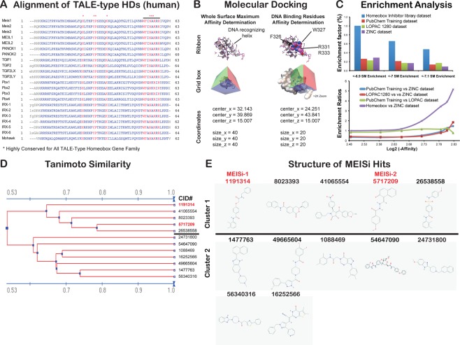
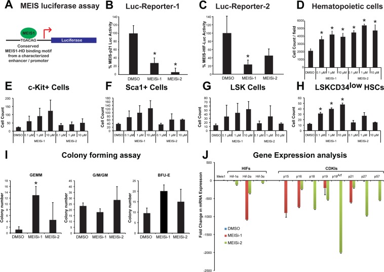
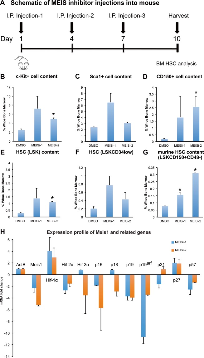
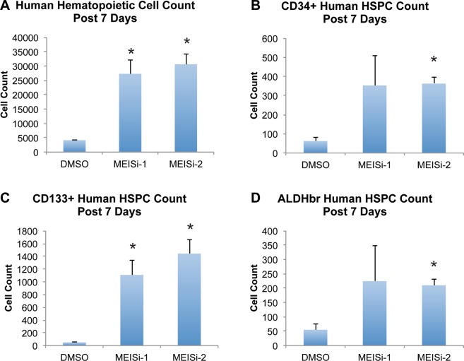
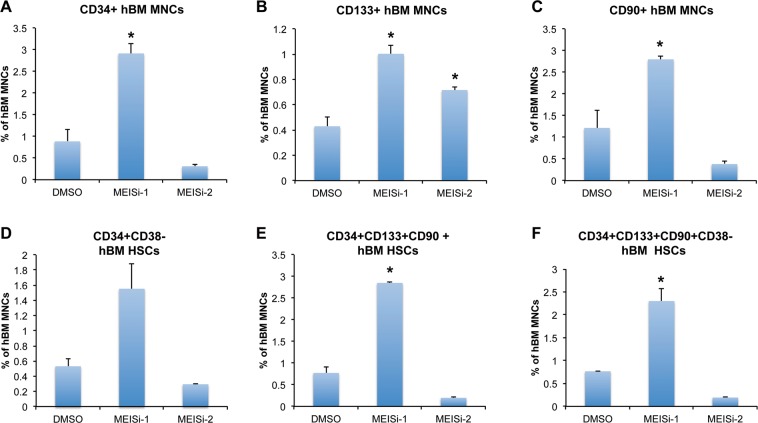
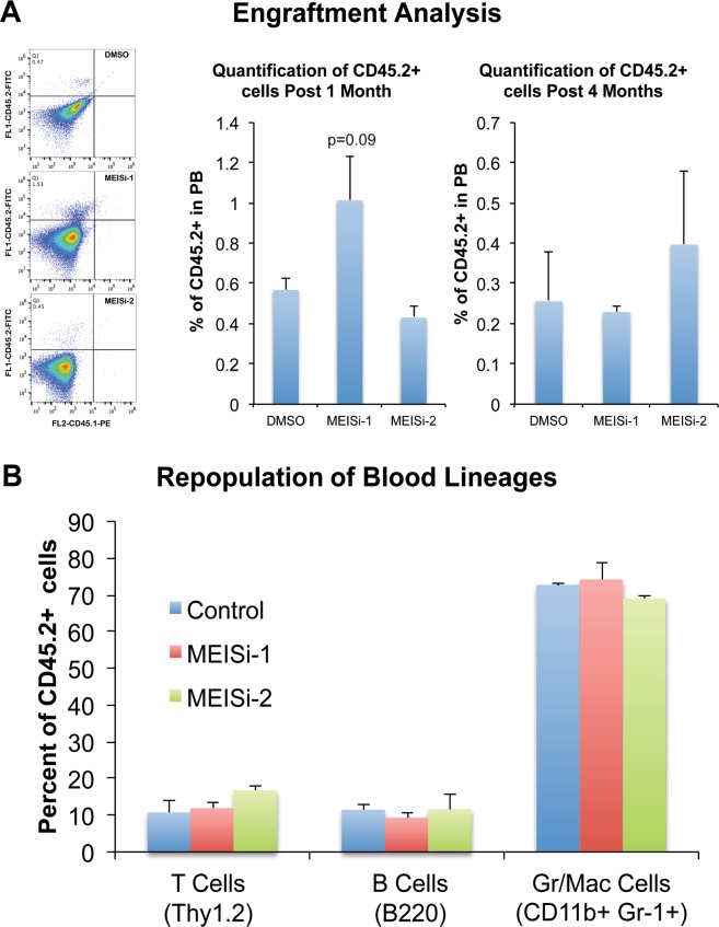
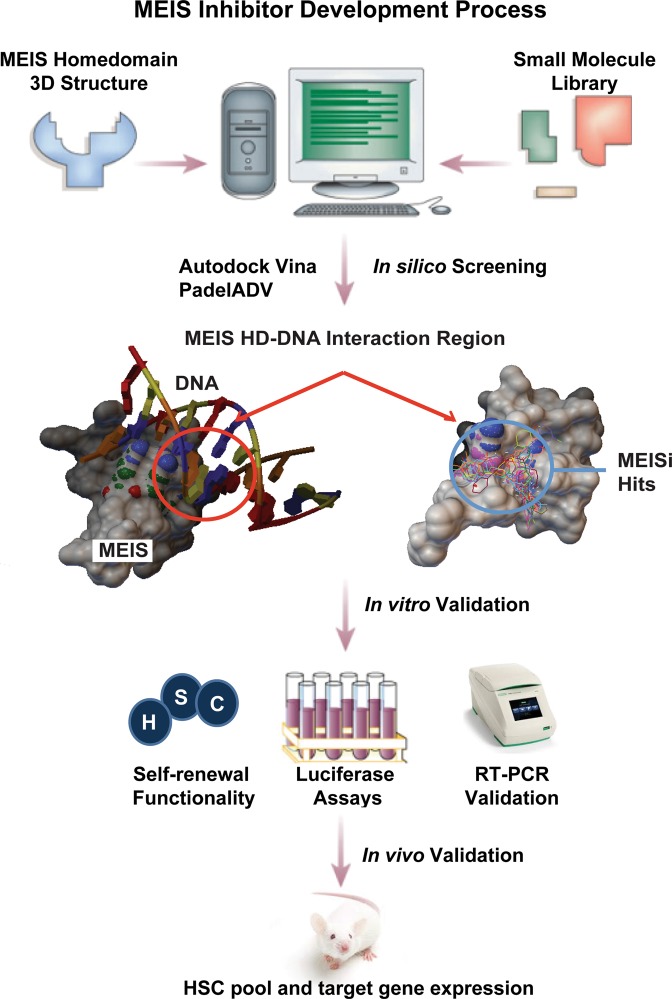
Meis1, which belongs to TALE-type class of homeobox gene family, appeared as one of the key regulators of hematopoietic stem cell (HSC) self-renewal and a potential therapeutical target. However, small molecule inhibitors of MEIS1 remained unknown. This led us to develop inhibitors of MEIS1 that could modulate HSC activity. To this end, we have established a library of relevant homeobox family inhibitors and developed a high-throughput in silico screening strategy against homeodomain of MEIS proteins using the AutoDock Vina and PaDEL-ADV platform. We have screened over a million druggable small molecules in silico and selected putative MEIS inhibitors (MEISi) with no predicted cytotoxicity or cardiotoxicity. This was followed by in vitro validation of putative MEIS inhibitors using MEIS dependent luciferase reporter assays and analysis in the ex vivo HSC assays. We have shown that small molecules named MEISi-1 and MEISi-2 significantly inhibit MEIS-luciferase reporters in vitro and induce murine (LSKCD34l°w cells) and human (CD34+, CD133+, and ALDHhi cells) HSC self-renewal ex vivo. In addition, inhibition of MEIS proteins results in downregulation of Meis1 and MEIS1 target gene expression including Hif-1α, Hif-2α and HSC quiescence modulators. MEIS inhibitors are effective in vivo as evident by induced HSC content in the murine bone marrow and downregulation of expression of MEIS target genes. These studies warrant identification of first-in-class MEIS inhibitors as potential pharmaceuticals to be utilized in modulation of HSC activity and bone marrow transplantation studies.


Conflict of interest statement

Dr. Fatih Kocabas is founder of Meinox Pharma Technologies. All other authors declare that they have no conflicts of interest concerning this work.

Figures

Figure 1
Figure 1
Development of MEIS-HD inhibitors. (A) Alignment of TALE type HDs. The amino acid sequences of each TALE family proteins collected from NCBI. Multiple alignments were done using the “constraint based multiple alignment tool” (COBALT). Highly conserved residues are marked with *. Note that there are several 100% conserved residues across TALE family of proteins. (B) Molecular docking of MEIS homeodomain. MEIS homeodomain was docked with small molecules in two grid boxes, whole surface included grid box and DNA binding residues grid box. Whole surface search box is performed to determine and eliminate small molecules non-specifically binds to homeodomain versus DNA binding residues. Dimensions in the grid box are provided in angstroms. (C) Enrichment analysis. Percent enrichment and enrichment factor ratio analysis of hits in homeobox library of small molecules docked into MEIS HD compared to random (ZINC drugs-now set) or unrelated (Lopac1280 set, or PubChem training set) libraries. (D) Analysis of MEISi hit small molecules. MEIS hit molecules analyzed for structural similarity based on substructure key-based 2D Tanimoto similarity. (E) Two clusters of MEISi hit molecules were identified. Structures and CID numbers are provided.
Figure 2
Figure 2
(A) MEIS luciferase reporter assay. Schematic showing luciferase reporter with MEIS binding motif TGACAG in the regulatory regions that is used to test activity of Meis1 protein. (B) MEIS-Luc-Reporter 1. Two small molecules named MEISi-1 and MEISi-2 demonstrated inhibition of MEIS-p21-luciferase reporter (luciferase reporter with well characterized MEIS1 binding motif from p21 regulatory region) up to 90% at 0.1 µM concentration. (C) MEIS-Luc-Reporter 2. MEISi-1 and MEISi-2 demonstrated inhibition of MEIS-HIF-luciferase reporter (luciferase reporter with well characterized MEIS1 binding motif from Hif-1α enhancer region). (D) Effect of MEISi treatments on the Lin- cells ex vivo. Lin- cells were isolated and treated with corresponding MEIS inhibitors and doses. Post 7 days of treatment, cells were stained with Hoechst 33342 and counted using automated cell imaging platform. Hematopoietic stem and progenitor cell expansion post MEISi treatments. Lin- cells were isolated and treated with corresponding MEIS inhibitors and doses. Post 7 days of treatment, HSCs were stained with corresponding surface antigens (E) c-Kit+, (F) Sca1+, (G) LSK and (H) LSKCD34low and determined HSC content by flow cytometry. (I) Colony forming assay. Lin- cells were treated with MEIS inhibitors for seven days. Then, methocult based CFU assays were performed. Types of colonies formed post 12 days were quantified and illustrated as CFU-GEMM, CFU-G/M/GM and BFU-E colonies. (J) Expression of MEIS target genes, HIFs and CDKIs post MEISi treatments in Lin- Cells. Lin- cells were treated in vitro with MEIS inhibitors and collected RNA post 3 days of treatment for analysis of gene expression. Note that MEIS1 is known to transcriptionally regulate expression of Hif-1α, Hif-2α, p15, p19ARF and p21. n = 3, *p < 0.05.
Figure 3
Figure 3
Phenotypical HSC antigen analysis post MEISi injections. (A) Schematic showing the intraperitoneal injections of MEIS inhibitors and BM analysis at day 10. In vivo analysis of HSC compartment post MEISi treatments was carried out by analysis of (B) c-Kit+, (C) Sca-1+ cell content, (D) CD150+ cell content, (E) LSK cell content, (F) LSKCD34low cell content and (G) LSKCD48CD150+ HSC content in the whole bone marrow following injection of MEISi-1, MEISi-2 and DMSO control. (H) Expression profile of Meis1 and related target genes post MEISi injections in the whole bone marrow cells. n = 3, *p < 0.05.
Figure 4
Figure 4
Human UCB hematopoietic cell expansion post MEISi treatments. (A) Human Hematopoietic cell count post 7 days of MEISi treatments, (B) CD34+ human HSPC count post 7 days, (C) CD133+ human HSPC count post 7 days, (D) ALDHbr human HSPC count post 7 days. n = 3. *p < 0.05.
Figure 5
Figure 5
Human bone marrow hematopoietic cell expansion post MEISi treatments. (A) the percentage of CD34+ hBM MNC content, (B) the percentage of CD133+ hBM MNC content, (C) the percentage of CD90+hBM MNC content D) the percentage of CD34+CD38 hBM MNC content, (E) the percentage of CD34+CD133+CD90+ hBM MNC content, (F) the percentage of CD34+CD133+CD90+CD38 hBM MNC post 7 days of treatment with MEISi-1 and MEISi-2 compared to the cells treated with DMSO. n = 3, *p < 0.05.
Figure 6
Figure 6
Analysis of engraftment and repopulation post MEISi expanded HSC transplantation. Murine LSK cells expressing CD45.2 allele were treated with effective dose of MEIS inhibitors and expanded for 7 days in HSC medium. These expanded HSCs were injected to NOD/SCID mice expressing CD45.1allele in a retro-orbital way. After 1 and 4 months, peripheral blood analysis was performed. Flow cytometry analysis was done with anti-CD45.2-FITCH and anti-CD45.1-PE. (A) Engraftment analysis. Engraftment and repopulation of HSCs were quantified based on the percentage of CD45.2+CD45.1 cells in the peripheral blood. (B) Repopulation of blood lineages. Quantification of B cells, granulocytes/macrophages and T cells derived from CD45.2+ HSCs in the recipient were determined. n = 4.
Figure 7
Figure 7
MEIS inhibitor development process. Targeting homeodomain of MEIS proteins and scoring against other TALE homeodomain proteins allowed to identify MEIS specific small molecules from in silico screening. We have targeted the MEIS-DNA interaction. This is followed by in vitro and in vivo validations. We have shown that small molecules named MEISi-1 and MEISi-2 inhibit MEIS-Luciferase reporters. Inhibition of MEIS proteins could functionally extend self-renewal of murine and human HSPCs ex vivo. Inhibition of MEIS proteins results in downregulation of Meis1, Meis2 and MEIS target genes including Hif-1α, Hif-2α and a number of HSC quiescence modulators. MEIS inhibitors are effective in vivo as evident by induced HSC content in the murine bone marrow and downregulation of expression of key MEIS target genes. MEIS inhibitors could be used to modulate human BM mPB HSC self-renewal as evident by increased number of CD34+CD38 cells.

References

    1. Cesselli D, et al. Oxidative stress-mediated cardiac cell death is a major determinant of ventricular dysfunction and failure in dog dilated cardiomyopathy. Circ Res. 2001;89:279–86. doi: 10.1161/hh1501.094115. - DOI - PubMed
    1. Argiropoulos B, Humphries RK. Hox genes in hematopoiesis and leukemogenesis. Oncogene. 2007;26:6766–76. doi: 10.1038/sj.onc.1210760. - DOI - PubMed
    1. Ariki R, et al. Homeodomain transcription factor Meis1 is a critical regulator of adult bone marrow hematopoiesis. PLoS One. 2014;9:e87646. doi: 10.1371/journal.pone.0087646. - DOI - PMC - PubMed
    1. von Burstin J, Reichert M, Wescott MP, Rustgi AK. The pancreatic and duodenal homeobox protein PDX-1 regulates the ductal specific keratin 19 through the degradation of MEIS1 and DNA binding. PLoS One. 2010;5:e12311. doi: 10.1371/journal.pone.0012311. - DOI - PMC - PubMed
    1. Aksoz M, Turan RD, Albayrak E, Kocabas F. Emerging Roles of Meis1 in Cardiac Regeneration, Stem Cells and Cancer. Curr Drug Targets. 2018;19:181–190. doi: 10.2174/1389450118666170724165514. - DOI - PubMed

Publication types

MeSH terms

Substances