

Fluorescence labeling of a NaV1.7-targeted peptide for near-infrared nerve visualization
- PMID: 32409881
- PMCID: PMC7225226
- DOI: 10.1186/s13550-020-00630-4
Fluorescence labeling of a NaV1.7-targeted peptide for near-infrared nerve visualization
Abstract
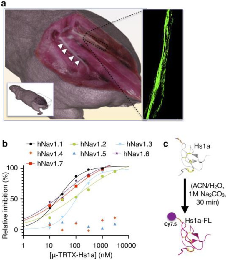
Background: Accidental peripheral nerve injury during surgical intervention results in a broad spectrum of potentially debilitating side effects. Tissue distortion and poor visibility can significantly increase the risk of nerve injury with long-lasting consequences for the patient. We developed and characterized Hs1a-FL, a fluorescent near-infrared molecule for nerve visualization in the operating theater with the aim of helping physicians to visualize nerves during surgery. Hs1a was derived from the venom of the Chinese bird spider, Haplopelma schmidti, and conjugated to Cy7.5 dye. Hs1a-FL was injected intravenously in mice, and harvested nerves were imaged microscopically and with epifluorescence.
Results: Hs1a-FL showed specific and stable binding to the sodium channel NaV1.7, present on the surface of human and mouse nerves. Hs1a-FL allowed epifluorescence visualization of sciatic mouse nerves with favorable nerve-to-muscle contrast.
Conclusions: Fluorescent NaV1.7-targeted tracers have the potential to be adopted clinically for the intraoperative visualization of peripheral nerves during surgery, providing guidance for the surgeon and potentially improving the standard of care.
Keywords: Hs1a-FL; Intraoperative; Near-infrared imaging; Nerve imaging.
Conflict of interest statement
All of the authors have read and approved the manuscript and possible conflict of interests are disclosed.
T.R. is shareholder of Summit Biomedical Imaging, LLC, and paid consultant for Theragnostics, Inc. J.G., P.D.d.S.F., G.F.K., J.S.L., and T.R. filed a patent surrounding the use of fluorophores with Hs1a and Hsp1a.
Figures


Similar articles
-
Development and Validation of Nerve-Targeted Bacteriochlorin Sensors.J Am Chem Soc. 2023 Jul 5;145(26):14276-14287. doi: 10.1021/jacs.3c02520. Epub 2023 Jun 20. J Am Chem Soc. 2023. PMID: 37339504 Free PMC article.
-
Fluorescence Imaging of Peripheral Nerves by a Nav1.7-Targeted Inhibitor Cystine Knot Peptide.Bioconjug Chem. 2019 Nov 20;30(11):2879-2888. doi: 10.1021/acs.bioconjchem.9b00612. Epub 2019 Nov 8. Bioconjug Chem. 2019. PMID: 31647222 Free PMC article.
-
NaV1.7 targeted fluorescence imaging agents for nerve identification during intraoperative procedures.bioRxiv [Preprint]. 2024 Apr 6:2024.04.06.588368. doi: 10.1101/2024.04.06.588368. bioRxiv. 2024. PMID: 38617358 Free PMC article. Preprint.
-
Fluorescence Imaging of Nerves During Surgery.Ann Surg. 2019 Jul;270(1):69-76. doi: 10.1097/SLA.0000000000003130. Ann Surg. 2019. PMID: 30649014
-
Structure-Inherent Targeting of Near-Infrared Fluorophores for Image-Guided Surgery.Chonnam Med J. 2017 May;53(2):95-102. doi: 10.4068/cmj.2017.53.2.95. Epub 2017 May 25. Chonnam Med J. 2017. PMID: 28584787 Free PMC article. Review.
Cited by
-
Pain-related toxins in scorpion and spider venoms: a face to face with ion channels.J Venom Anim Toxins Incl Trop Dis. 2021 Dec 6;27:e20210026. doi: 10.1590/1678-9199-JVATITD-2021-0026. eCollection 2021. J Venom Anim Toxins Incl Trop Dis. 2021. PMID: 34925480 Free PMC article. Review.
-
Realizing real-time optical molecular imaging in peripheral nerve tissue via Rhodamine B.Front Med (Lausanne). 2024 Nov 26;11:1461520. doi: 10.3389/fmed.2024.1461520. eCollection 2024. Front Med (Lausanne). 2024. PMID: 39659623 Free PMC article.
-
Development and Validation of Nerve-Targeted Bacteriochlorin Sensors.J Am Chem Soc. 2023 Jul 5;145(26):14276-14287. doi: 10.1021/jacs.3c02520. Epub 2023 Jun 20. J Am Chem Soc. 2023. PMID: 37339504 Free PMC article.
-
Tissue-seeking dyes for in vivo applications.Smart Mol. 2024 Oct 24;2(4):e20240029. doi: 10.1002/smo.20240029. eCollection 2024 Dec. Smart Mol. 2024. PMID: 40626275 Free PMC article. Review.
-
Chemical and Biological Tools for the Study of Voltage-Gated Sodium Channels in Electrogenesis and Nociception.Chembiochem. 2022 Jul 5;23(13):e202100625. doi: 10.1002/cbic.202100625. Epub 2022 Mar 21. Chembiochem. 2022. PMID: 35315190 Free PMC article. Review.
References
Grants and funding
LinkOut - more resources
Full Text Sources
Other Literature Sources
