Review
. 2020 Dec;9(2):213-227.
doi: 10.1007/s40120-020-00190-8. Epub 2020 May 14.

Pathophysiology of Chronic Inflammatory Demyelinating Polyneuropathy: Insights into Classification and Therapeutic Strategy

Affiliations
Review

Pathophysiology of Chronic Inflammatory Demyelinating Polyneuropathy: Insights into Classification and Therapeutic Strategy

Haruki Koike et al. Neurol Ther. 2020 Dec.

Abstract

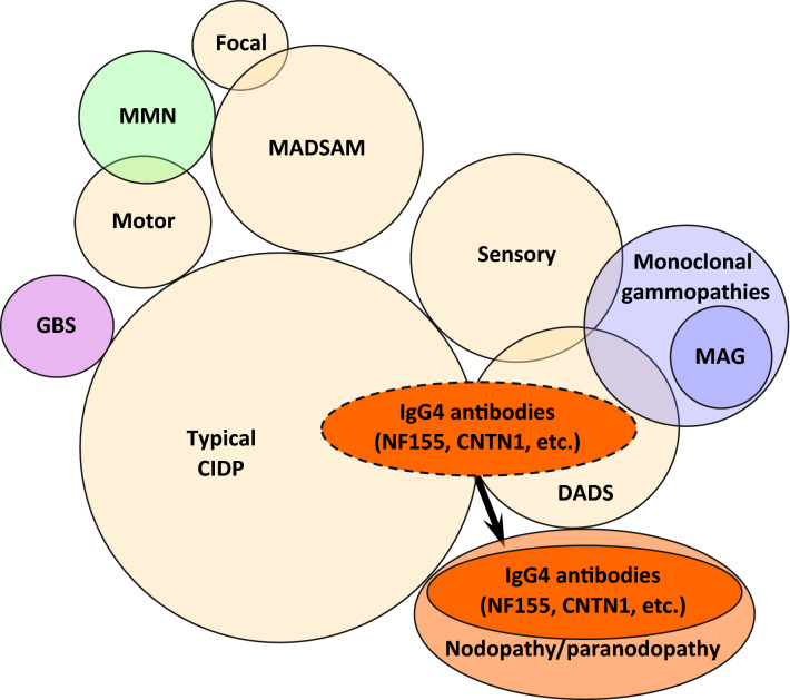
Chronic inflammatory demyelinating polyneuropathy (CIDP) is classically defined as polyneuropathy with symmetric involvement of the proximal and distal portions of the limbs. In addition to this "typical CIDP", the currently prevailing diagnostic criteria proposed by the European Federation of Neurological Societies and Peripheral Nerve Society (EFNS/PNS) define "atypical CIDP" as encompassing the multifocal acquired demyelinating sensory and motor (MADSAM), distal acquired demyelinating symmetric (DADS), pure sensory, pure motor, and focal subtypes. Although macrophage-induced demyelination is considered pivotal to the pathogenesis of CIDP, recent studies have indicated the presence of distinctive mechanisms initiated by autoantibodies against paranodal junction proteins, such as neurofascin 155 and contactin 1. These findings led to the emergence of the concept of nodopathy or paranodopathy. Patients with these antibodies tend to show clinical features compatible with typical CIDP or DADS, particularly the latter. In contrast, classical macrophage-induced demyelination is commonly found in some patients in each major subtype, including the typical CIDP, DADS, MADSAM, and pure sensory subtypes. Differences in the distribution of lesions and the repair processes underlying demyelination by Schwann cells may determine the differences among subtypes. In particular, the preferential involvement of proximal and distal nerve segments has been suggested to occur in typical CIDP, whereas the involvement of the middle nerve segments is conspicuous in MADSAM. These findings suggest that humoral rather than cellular immunity predominates in the former because nerve roots and neuromuscular junctions lack blood-nerve barriers. Treatment for CIDP consists of intravenous immunoglobulin (IVIg) therapy, steroids, and plasma exchange, either alone or in combination. However, patients with anti-neurofascin 155 and contactin 1 antibodies are refractory to IVIg. It has been suggested that rituximab, a monoclonal antibody to CD20, could have efficacy in these patients. Further studies are needed to validate the CIDP subtypes defined by the EFNS/PNS from the viewpoint of pathogenesis and establish therapeutic strategies based on the pathophysiologies specific to each subtype.

Keywords: Demyelination; Electron microscopy; Macrophage; Node of Ranvier; Paranode; Pathogenesis; Pathology; Schwann cell; Treatment; Ultrastructure.


Conflict of interest statement

Haruki Koike and Masahisa Katsuno have nothing to disclose.

Figures

Fig. 1
Fig. 1
Representative photographs of macrophage-induced demyelination. Sural nerve biopsy specimens obtained from a patient with typical CIDP. a Teased-fiber preparations showing segments devoid of myelin (indicated by arrows) as a result of phagocytosis by macrophages. b Cross sections of epoxy-resin-embedded specimens showing macrophages surrounding myelinated fibers (arrow). c Electron microscopy showing that these macrophages contain myelin debris in their cytoplasm. A high-powered view of the region shown in the box in c is shown in d. d Layers of myelin lamellae apposed to the cytoplasm of macrophages become fuzzy as a result of the disruption of myelin lamellae. Osmium stain (a), toluidine blue stain (b), and uranyl acetate and lead citrate stain (c, d). Scale bars 50 μm (a), 10 μm (b), 2 μm (c), and 0.2 μm (d)
Fig. 2
Fig. 2
Representative electron microscopy photograph of paranodal dissection. Sural nerve biopsy specimens obtained from a patient with anti-neurofascin 155 antibodies (a) and a control subject (b). Longitudinal sections. a Clear spaces are shown between the myelin terminal loops and axolemma (arrows). b Normally, the terminal loops of myelin are closely apposed to the axolemma at paranodes. Uranyl acetate and lead citrate stain. Scale bars 0.5 μm
Fig. 3
Fig. 3
Representative pathological findings in patients with atypical CIDP. Sural nerve biopsy specimens obtained from patients with pure sensory (a, c) and MADSAM (multifocal acquired demyelinating sensory and motor) (b). Transverse sections. a Conspicuous variation in myelinated fiber density among fascicles. b Mild onion bulb formation in areas of reduced myelinated fiber density in a patient with MADSAM. c In a patient with pure sensory subtype, the reduction in myelinated fiber density seemed to result from an enlargement of the cross-sectional area due to marked onion bulb formation. Toluidine blue staining (a) and uranyl acetate and lead citrate staining (b, c). Scale bars 50 μm (a), 1 μm (b), and 10 μm (c)
Fig. 4
Fig. 4
A correlation diagram of CIDP subtypes defined in the EFNS/PNS criteria and their related diseases. Patients with IgG4 antibodies to paranodal neurofascin 155 and contactin 1 show mechanisms of neuropathy distinct from those observed in classical macrophage-induced demyelination, indicating that specific therapeutic strategies are involved in these two groups of patients. In particular, the concept of nodopathy or paranodopathy has recently been proposed for patients with IgG4 antibodies to nodal and paranodal components. Hence, clarifying whether the classification of this group of patients as “CIDP” is appropriate is a challenge for the future. CNTN1 contactin 1, DADS distal acquired demyelinating symmetric, GBS Guillain–Barré syndrome, MADSAM multifocal acquired demyelinating sensory and motor, MAG myelin-associated glycoprotein, MMN multifocal motor neuropathy, Motor pure motor, NF155 neurofascin 155, Sensory pure sensory

References

    1. Dyck PJ, Lais AC, Ohta M, Bastron JA, Okazaki H, Groover RV. Chronic inflammatory polyradiculoneuropathy. Mayo Clin Proc. 1975;50:621–637. - PubMed
    1. McCombe PA, Pollard JD, McLeod JG. Chronic inflammatory demyelinating polyradiculoneuropathy. A clinical and electrophysiological study of 92 cases. Brain. 1987;110:1617–1630. doi: 10.1093/brain/110.6.1617. - DOI - PubMed
    1. Barohn RJ, Kissel JT, Warmolts JR, et al. Chronic inflammatory demyelinating polyradiculoneuropathy. Clinical characteristics, course, and recommendations for diagnostic criteria. Arch Neurol. 1989;46:878–884. doi: 10.1001/archneur.1989.00520440064022. - DOI - PubMed
    1. Bouchard C, Lacroix C, Planté V, et al. Clinicopathologic findings and prognosis of chronic inflammatory demyelinating polyneuropathy. Neurology. 1999;52(3):498–503. doi: 10.1212/WNL.52.3.498. - DOI - PubMed
    1. Vallat JM, Sommer C, Magy L. Chronic inflammatory demyelinating polyradiculoneuropathy: diagnostic and therapeutic challenges for a treatable condition. Lancet Neurol. 2010;9:402–412. doi: 10.1016/S1474-4422(10)70041-7. - DOI - PubMed

LinkOut - more resources