

Toxoplasma gondii and Neospora caninum infections in South American camelids in Switzerland and assessment of serological tests for diagnosis
- PMID: 32410682
- PMCID: PMC7227098
- DOI: 10.1186/s13071-020-04128-9
Toxoplasma gondii and Neospora caninum infections in South American camelids in Switzerland and assessment of serological tests for diagnosis
Abstract
Background: Little is known about the epidemiology of Toxoplasma gondii and Neospora caninum infections in alpacas (Vicugna pacos) and llamas (Lama glama) outside South America. The study aimed to estimate the seroprevalence of T. gondii and N. caninum infections in South American camelids (SAC) in Switzerland, to optimize serological tests for SAC and to identify risk factors, which may favour infection.
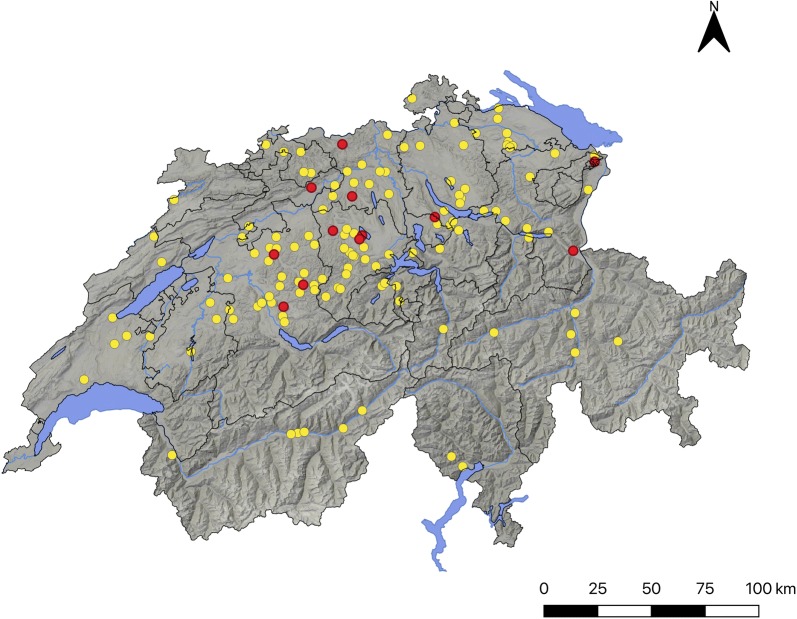
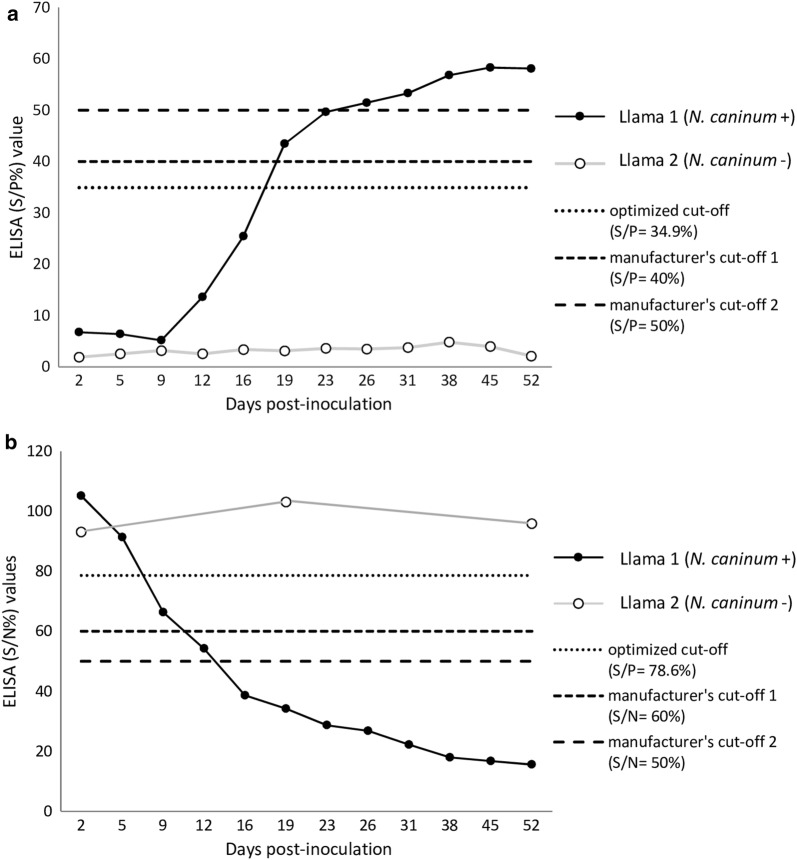
Methods: A total of 571 sera from 132 Swiss farms (374 alpacas and 197 llamas, mean 4.3 animals/farm) were obtained. Four commercial enzyme-linked immunosorbent assays (ELISA) for detecting antibodies against T. gondii (ID Screen® Toxoplasmosis Indirect (TOXO-MS)) or N. caninum (i.e. ID Screen® Neospora caninum Indirect Multi-species (NCS-MS); ID Screen® Neospora caninum Competition (NCC) and ID Screen® Neospora caninum Indirect (NCS)) were first assessed for their use on SAC comparing their results with those in immunoblot, and optimizing cut-offs. Subsequently, two kits (TOXO-MS and NCS-MS) were selected for seroprevalence estimation. Additionally, a risk factor analysis for infection was performed on 41 farms, which agreed to participate in a web-based survey.
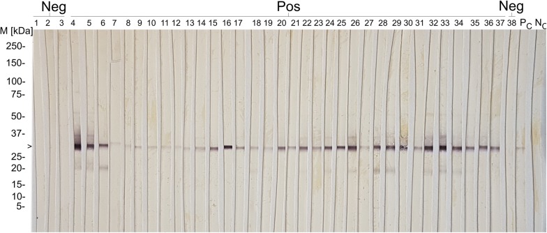
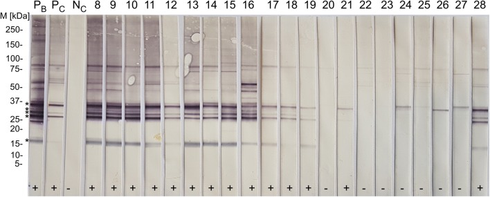
Results: Three kits (TOXO-MS, NCS-MS and NCC) showed almost perfect agreement (kappa > 0.901) with immunoblot results when the cut-offs were optimized, and one kit (NCS) proved not to be useful for detecting N. caninum seropositive SAC. By TOXO-MS ELISA, 82.3% (308/374) of the alpacas and 84.8% (167/197) of the llamas were seropositive for T. gondii, and 131/132 (99.2%) farms had seropositive animals. By NCS-MS ELISA, 3.5% (13/374) of the alpacas and 2.5% (5/197) of the llamas evidenced antibodies against N. caninum, and 9.1% (12/132) of the farms had seropositive animals. The variables "age" and "female sex" were identified as risk factors for T. gondii infection and "absence of cats in the farm during the last two years" as a protective factor. No risk or protective factors for N. caninum infection could be identified.
Conclusions: This nationwide cross-sectional study demonstrated for the first time the presence of antibodies against T. gondii and N. caninum in the Swiss SAC population, highlighting a high seroprevalence for T. gondii, the presence of cats as a risk factor and suggesting that SAC meat might represent an additional infection source for humans.
Keywords: Alpaca; ELISA; Immunoblot; Lama glama; Llama; Neosporosis; Toxoplasmosis; Vicugna pacos.
Conflict of interest statement
The authors declare that they have no competing interests.
Figures
References
-
- Dubey JP. Toxoplasmosis of animals and humans. 2. Boca Raton: CRC Press; 2010.
-
- Dubey JP, Hemphill A, Calero-Bernal R, Schares G. Neosporosis in animals. Portland: CRC Press; 2017.
-
- Broglia A, Basso W. Parasites present in meat and viscera of land farmed animals. In: Devine C, Dikeman M, editors. Encyclopedia of meat sciences 2e. Oxford: Elsevier; 2014. pp. 34–41.
-
- Basso W, Edelhofer R, Zenker W, Möstl K, Kübber-Heiss A, Prosl H. Toxoplasmosis in Pallasʼ cats (Otocolobus manul) raised in captivity. Parasitology. 2005;130:293–299. - PubMed
-
- Basso W, Moré G, Quiroga MA, Pardini L, Bacigalupe D, Venturini L, et al. Isolation and molecular characterization of Toxoplasma gondii from captive slender-tailed meerkats (Suricata suricatta) with fatal toxoplasmosis in Argentina. Vet Parasitol. 2009;161:201–206. - PubMed
MeSH terms
Substances
LinkOut - more resources
Full Text Sources
Miscellaneous
