. 2020 Apr 7;17(8):1030-1042.
doi: 10.7150/ijms.43986. eCollection 2020.

Fibroblast Proliferation and Migration in Wound Healing by Phytochemicals: Evidence for a Novel Synergic Outcome

Affiliations

Fibroblast Proliferation and Migration in Wound Healing by Phytochemicals: Evidence for a Novel Synergic Outcome

Roberta Addis et al. Int J Med Sci. .

Abstract

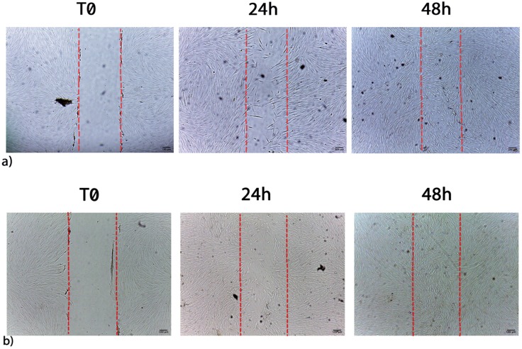
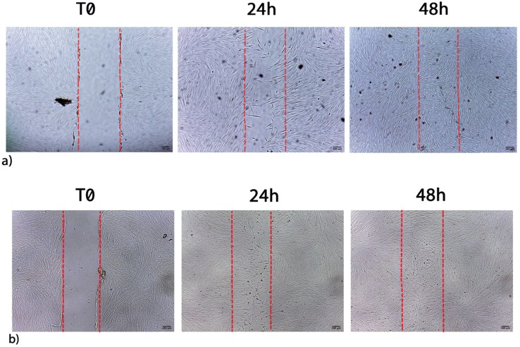
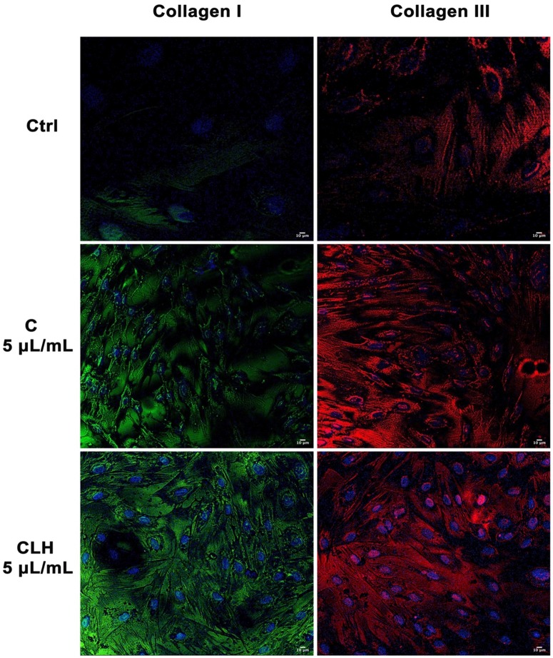
Wound-healing is a dynamic skin reparative process that results in a sequence of events, including inflammation, proliferation, and migration of different cell types as fibroblasts. Fibroblasts play a crucial role in repairing processes, from the late inflammatory phase until the fully final epithelization of the injured tissue. Within this context, identifying tools able to implement cell proliferation and migration could improve tissue regeneration. Recently, plants species from all over the world are coming out as novel tools for therapeutic applications thanks to their phytochemicals, which have antioxidant properties and can promote wound healing. In this paper, we aimed at investigating antioxidant activity of waste extracts from different medicinal plants, endemic of the Mediterranean area, on fibroblast proliferation and wound healing. We determined the amount of total phenols and anti-oxidant activity by ABTS assay. We then evaluated the cytotoxicity of the compounds and the proliferative capabilities of fibroblasts by scratch assay. Our results showed that waste extracts retain antioxidant and regenerative properties, inducing tissue re-establishment after environmental stress exposure. Taken together, our findings suggest that waste material could be used in the future also in combinations to stimulate wound healing processes and antioxidant responses in damaged skin.

Keywords: antioxidants; cell proliferation; cellular mechanisms; natural molecules; oxidative stress; tissue regeneration.


Conflict of interest statement

Competing Interests: The authors have declared that no competing interest exists.

Figures

Figure 1
Figure 1
(a) Linear regression analysis of phenols amounts of waste extracts of C. arvensis (C), L. stoechas (L), and H. italicum (H) and (b) pooled waste extracts of C. arvensis, L. stoechas, and H. italicum (CLH, CL, CH, LH) over volumes. Data were expressed as mean of 3 independent experiments (*P<0.05; **P<0.01).
Figure 2
Figure 2
Time-course of antioxidant activity of waste extracts from leaves of C. arvensis (C), L. stoechas (L), and H. italicum (H) and pooled waste extracts (CLH, CL, CH, LH) evaluated by ABTS assays. (a) Free radical scavenging of 50% of ABTS by Trolox, used as standard, C or L or H at the indicated time-points. The values are expressed as mean ± SD of three independent assays (*P < 0.05). (b) Free radical scavenging of 50% of ABTS by Trolox, and pooled waste extracts CLH, CL, CH, LH at the indicated time points. The values are expressed as mean ± SD of three independent assays (*P < 0.05).
Figure 3
Figure 3
MTT assay of waste extract of C. arvensis (C), L. stoechas (L), and H. italicum (H) at different concentration (1, 5, 10 μL/mL) after 24h (a), 48h (b), 72h (c), 96h (d) and 120h (e) (* p<0.05 ** p<0.01 as compared to untreated control (0).
Figure 4
Figure 4
MTT assay of different concentration (1, 5, 10 μL/mL) of pooled waste extracts of C. arvensis, L. stoechas, and H. italicum CLH (light blue bar), CL (pink bar), CH (purple bar), LH (grey bar), after 24h (a), 48h (b), 72h (c), 96h (d) and 120h (e) (* p<0.05 as compared to untreated control (0).
Figure 5
Figure 5
a) Migration of control cells after scratch. b) Migration of fibroblasts after scratch and treatment with 1 µL/mL L extracts. Images are acquired by optical microscope and are examples of different independent experiments. Scale bar = 100 µm.
Figure 6
Figure 6
a) Migration of control cells after scratch. b) Migration of fibroblasts after scratch and treatment with 1 μL/mL CLH extracts. Images are acquired by optical microscope and are examples of different independent experiments. Scale bar = 100 µm.
Figure 7
Figure 7
Analysis of collagen deposition during wound healing. Immunohistochemical analysis of the expression of Collagen type I and type III was assessed in fibroblasts after scratch and treatment with different concentrations of single waste extract (C, L, H) or pooled waste extracts (CLH, CL, CH, LH) for 72 hours. Control cells were cultured in basic growing medium. Nuclei are labelled with 4,6-diamidino-2-phenylindole (DAPI, blue). Scale bars: 10 µm. The figures are representative of different independent experiments. For each differentiation marker, fields with the highest yield of positively stained cells are shown.

References

    1. Barrientos S, Stojadinovic O, Golinko MS. et al. Growth factors and cytokines in wound healing. Wound Repair and Regeneration. 2008;(5):585–601. - PubMed
    1. Behm D, Colado JC. The effectiveness of resistance training using unstable surfaces and devices for rehabilitation. Int. J. Sports Phys. Ther. 2012;7(2):226–41. - PMC - PubMed
    1. Maddaluno L, Urwyler C, Werner S. Fibroblast growth factors: Key players in regeneration and tissue repair. Development (Cambridge) 2017;144(22):4047–4060. - PubMed
    1. Bainbridge P. Wound healing and the role of fibroblasts. J. Wound Care. 2013;22:407. - PubMed
    1. Tracy LE, Minasian RA, Caterson EJ. Extracellular Matrix and Dermal Fibroblast Function in the Healing Wound. Advances in Wound Care. 2016;(3):119–136. - PMC - PubMed