. 2020 Jan 24:2020:7649480.
doi: 10.1155/2020/7649480. eCollection 2020.

The Role of Angiogenesis Factors in the Formation of Vascular Changes in Scleroderma by Assessment of the Concentrations of VEGF and sVEGFR2 in Blood Serum and Tear Fluid

Affiliations

The Role of Angiogenesis Factors in the Formation of Vascular Changes in Scleroderma by Assessment of the Concentrations of VEGF and sVEGFR2 in Blood Serum and Tear Fluid

Arleta Waszczykowska et al. Mediators Inflamm. .

Abstract

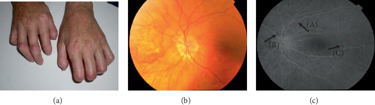
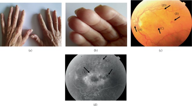
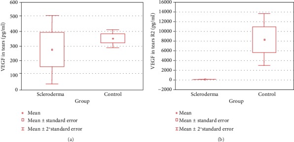
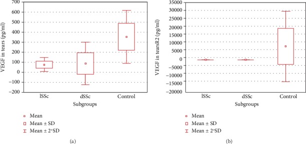
Systemic sclerosis (SSc) is a connective tissue disorder characterized by tissue hypoxia, excessive fibrosis of skin and internal organs, and angiogenesis imbalance. The aim of the study was to evaluate in SSc patients the association between the retinal microcirculation disturbances and the presence of peripheral trophic changes and to determine the role of angiogenesis factors in the formation of vascular changes in scleroderma. Twenty-five SSc patients and 25 age- and sex-matched healthy controls were included to the study. Assay of vascular endothelial growth factor (VEGF) and soluble VEGF receptor-2 (sVEGFR-2) in blood serum and tears was done for all patients and controls using enzyme-linked immunosorbent assay. Retinal blood circulation was investigated with fluorescein angiography (FA) in the SSc patients only. In our research, proportion of mainly hypertensive patients presenting with a large spectrum of retinal microvascular lesions was 72%, while proportion of patients with skin microvascular lesions within distal phalanxes of fingers and toes was 76%. We noticed that patients with pathological changes in the FA examination had finger ulcerations significantly more often than patients without changes in the eye fundus. There were no statistically significant differences in the serum concentration of VEGF and sVEGFR2 between subjects in both analyzed groups. Analysis of lower levels of VEGF (p = <0.001) and sVEGFR-2 (p = <0.001) in blood serum accompanied by simultaneous higher levels of VEGF/sVEGFR-2 ratio in tears of SSc patients, as compared with the control group, indicates the superiority of proangiogenic factors in patients' tears.


Conflict of interest statement

The authors report no conflict of interest.

Figures

Figure 1
Figure 1
Three images of hands (a) and the eye fundus (b, c) of a patient, aged 51, diagnosed with systemic sclerosis six years ago. Vascular changes in the form of lysis of the distal phalanges of the hands fingers are visible (a). Color photo of the left eye fundus (B): sectional narrowing of retinal vessels (A), choroidal vessel transgression due to thinning of choriocapillaries and retinal pigment epithelium (B) can be observed. Results of fluorescein angiography of the left eye (c). Irregularities in the course and caliber of the retinal vessels (A) and foci of fluorescence blockade with local pigment epithelium regrouping (B, C) may be noted.
Figure 2
Figure 2
Panel of four images of hands (a, b) and eye fundus (c, d) of a patient, aged 61, diagnosed with systemic sclerosis seven years previously. Puffy fingers can be observed on both hands (a). Small scars from healed vascular ulcers within the second and third fingertips of the right hand at the edge of the nail (b). Color photo of the left eye fundus with ischemic retinopathy (c). Vasodilation of the optic disc (A), intraretinal petechiae (B), diffuse macular edema (C), and scars after laser therapy (D) may be noted. Results of fluorescein angiography of the left eye (d). A diffuse, poorly circumscribed and intensive hyperfluorescence of the whole posterior pole was observed in the recirculation phase; this was consistent with diffuse macular edema (A). Hypofluorescence foci (B) may be noted in the projection of extravasations and post-laser foci within temporal arcades (C).
Figure 3
Figure 3
Box-and-whiskers plot of VEGF (a) and sVEGFR-2 (b) in patients with scleroderma and controls. Abbreviations: VEGF: vascular endothelial growth factor; sVEGFR-2: soluble form of vascular endothelial growth factor receptor 2; SD: standard deviation.
Figure 4
Figure 4
Box-and-whiskers plot of VEGF (a) and sVEGFR-2 (b) in patients with scleroderma subtypes and control. For the VEGF, three outliers were removed for the sake of plot clarity. Abbreviations: VEGF: vascular endothelial growth factor; sVEGFR-2: soluble form of vascular endothelial growth factor receptor 2; SD: standard deviation.

References

    1. Nadashkevich O., Davis P., Fritzler M. J. A proposal of criteria for the classification of systemic sclerosis. Medical Science Monitor. 2004;10:615–621. - PubMed
    1. Sahin H., Wasmuth H. E. Chemokines in tissue fibrosis. Biochimica et Biophysica Acta (BBA) - Molecular Basis of Disease. 2013;1832(7):1041–1048. doi: 10.1016/j.bbadis.2012.11.004. - DOI - PubMed
    1. Smith G., McLeod D., Foreman D., Boulton M. Immunolocalisation of the VEGF receptors FLT-1, KDR, and FLT-4 in diabetic retinopathy. British Journal of Ophthalmology. 1999;83(4):486–494. doi: 10.1136/bjo.83.4.486. - DOI - PMC - PubMed
    1. Veeravagu A., Hsu A. R., Cai W., Hou L. C., Tse V. C., Chen X. Vascular endothelial growth factor and vascular endothelial growth factor receptor inhibitors as anti-angiogenic agents in cancer therapy. Recent Patents on Anti-Cancer Drug Discovery. 2007;2(1):59–71. doi: 10.2174/157489207779561426. - DOI - PubMed
    1. Barleon B., Reusch P., Totzke F., et al. Soluble VEGFR-1 secreted by endothelial cells and monocytes is present in human serum and plasma from healthy donors. Angiogenesis. 2001;4(2):143–154. doi: 10.1023/a:1012245307884. - DOI - PubMed

MeSH terms

Substances