Review
. 2020 Apr 29:14:363.
doi: 10.3389/fnins.2020.00363. eCollection 2020.

Cortical Circuit Dysfunction as a Potential Driver of Amyotrophic Lateral Sclerosis

Affiliations
Review

Cortical Circuit Dysfunction as a Potential Driver of Amyotrophic Lateral Sclerosis

Aurore Brunet et al. Front Neurosci. .

Abstract

Amyotrophic lateral sclerosis (ALS) is a devastating neurodegenerative disease that affects selected cortical and spinal neuronal populations, leading to progressive paralysis and death. A growing body of evidences suggests that the disease may originate in the cerebral cortex and propagate in a corticofugal manner. In particular, transcranial magnetic stimulation studies revealed that ALS patients present with early cortical hyperexcitability arising from a combination of increased excitability and decreased inhibition. Here, we discuss the possibility that initial cortical circuit dysfunction might act as the main driver of ALS onset and progression, and review recent functional, imaging and transcriptomic studies conducted on ALS patients, along with electrophysiological, pathological and transcriptomic studies on animal and cellular models of the disease, in order to evaluate the potential cellular and molecular origins of cortical hyperexcitability in ALS.

Keywords: amyotrophic lateral sclerosis; cerebral cortex; extrinsic; hyperexcitability; intrinsic; network dysfunction.


Figures

FIGURE 1
FIGURE 1
Schematic representation of transcranial magnetic stimulation and cortical hyperexcitability in ALS. Transcranial magnetic stimulation (TMS) consists in the application of a magnetic field above the motor cortex, and the recording of motor evoked potentials in the abductor pollicis brevis muscle of the hand (left). This technique showed reduction or absence of parameters related to cortical inhibition: (right) short interval intracortical inhibition (SICI), cortical silent period (CSP) and long interval intracortical inhibition (LICI), and an increase in parameters related to cortical excitation: intracortical facilitation (ICF) and short interval intracortical facilitation (SICF).
FIGURE 2
FIGURE 2
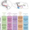
Schematic representation of the monoaminergic and cholinergic systems and their impairments in ALS patients and mouse models. Some monoamine and acetylcholine nuclei send projections to the motor cortex both in human (top left) and mouse brain (top right). Alterations in these systems have been found both in ALS patients (table, upper part) and ALS mouse models (table, lower part). Dorsal raphe nucleus and its serotonine (5-HT) projections are represented in green; ventral tegmental area (VTA) and dopamine (DA) projections in pink; locus coeruleus (LC) and noradrenaline (NA) projections in purple; tuberomammillary nucleus (TMN) and histamine projections in orange and basal forebrain (BF) and acetylcholine (ACh) projections in blue.
FIGURE 3
FIGURE 3
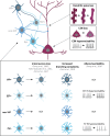
Schematic representation of the corticospinal neurons and GABAergic interneurons, and their alterations in ALS patients or mouse models. Corticospinal neurons (CSN) are excitatory glutamatergic neurons (dark pink) which, in ALS, present progressive dendritic spine loss (top right), cell loss and hyperexcitability of the remaining neurons (middle right). GABAergic interneurons (IN) are inhibitory neurons (four shades of blue) which can be classified in four groups: vasointestinal peptide (VIP) IN, somatostatin (SST) IN, non-VIP IN and Parvalbumin (PV) IN. Some alterations in IN density, branching complexity and excitability have been found in different ALS mouse models and ALS patients (bottom part).

References

    1. Agosta F., Spinelli E. G., Marjanovic I. V., Stevic Z., Pagani E., Valsasina P., et al. (2018). Unraveling ALS due to SOD1 mutation through the combination of brain and cervical cord MRI. Neurology 90 e707–e716. 10.1212/WNL.0000000000005002 - DOI - PubMed
    1. Agster K. L., Mejias-Aponte C. A., Clark B. D., Waterhouse B. D. (2013). Evidence for a regional specificity in the density and distribution of noradrenergic varicosities in rat cortex. J. Comp. Neurol. 521 2195–2207. 10.1002/cne.23270 - DOI - PMC - PubMed
    1. Albéri L., Lintas A., Kretz R., Schwaller B., Villa A. E. P. (2013). The calcium-binding protein parvalbumin modulates the firing 1 properties of the reticular thalamic nucleus bursting neurons. J. Neurophysiol. 109 2827–2841. 10.1152/jn.00072.2012 - DOI - PubMed
    1. Alberico S. L., Cassell M. D., Narayanan N. S. (2015). The vulnerable ventral tegmental area in Parkinson’s disease. Basal Ganglia. 5 51–55. 10.1016/j.baga.2015.06.001 - DOI - PMC - PubMed
    1. Andrés-Benito P., Moreno J., Aso E., Povedano M., Ferrer I. (2017). Amyotrophic lateral sclerosis, gene deregulation in the anterior horn of the spinal cord and frontal cortex area 8: implications in frontotemporal lobar degeneration. Aging 9 823–851. 10.18632/aging.101195 - DOI - PMC - PubMed