. 2020 Jan 9:2020:9207279.
doi: 10.1155/2020/9207279. eCollection 2020.

Lessons Learned from Experimental Human Model of Zinc Deficiency

Affiliations

Lessons Learned from Experimental Human Model of Zinc Deficiency

Ananda S Prasad. J Immunol Res. .

Abstract

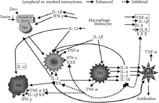
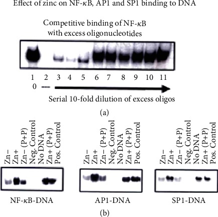
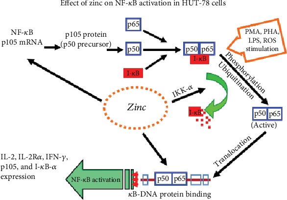
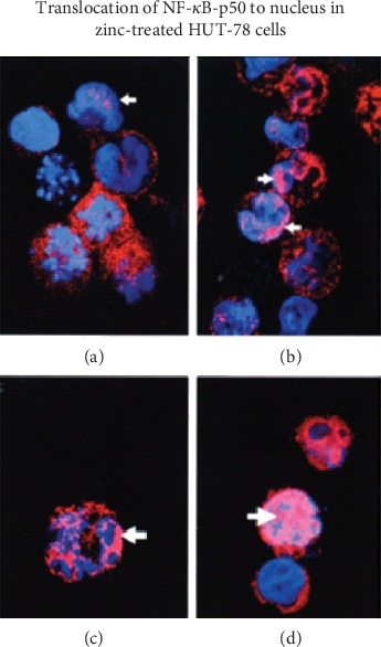
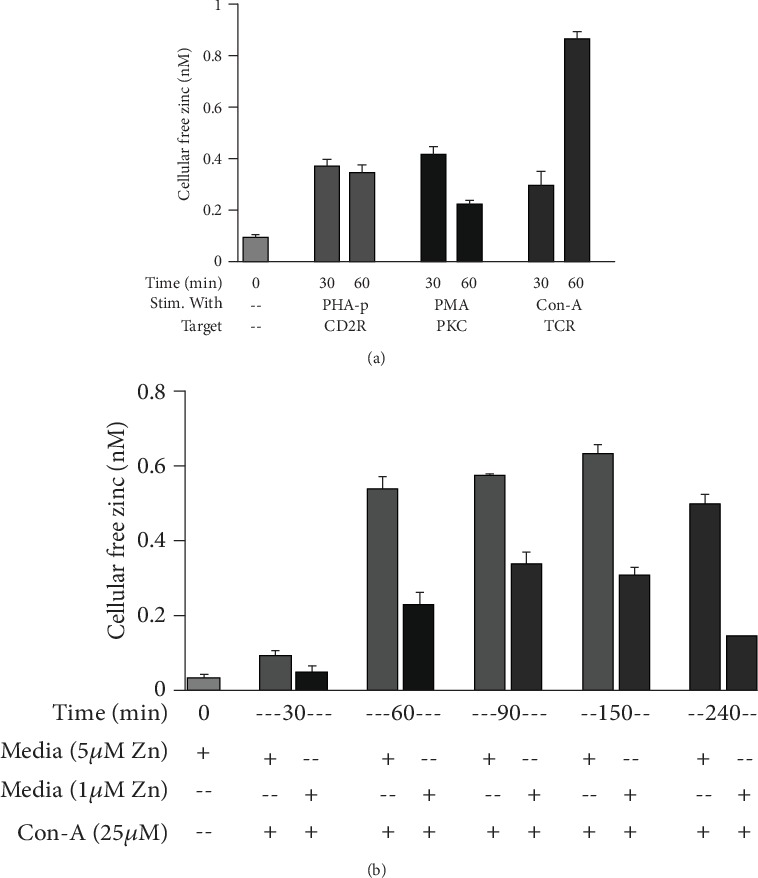
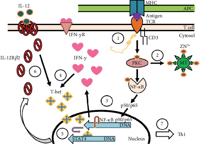
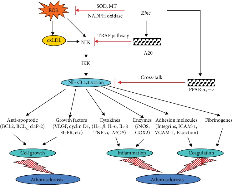
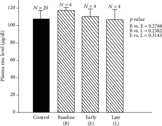
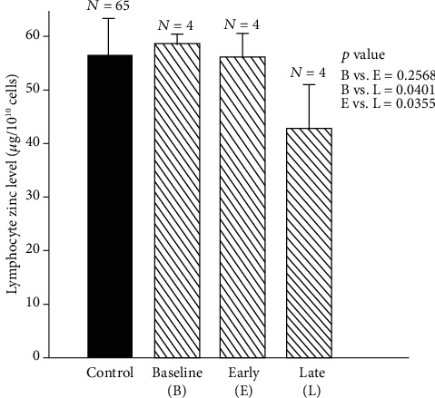
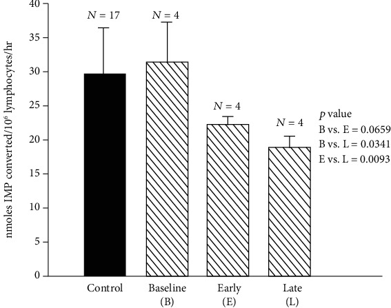
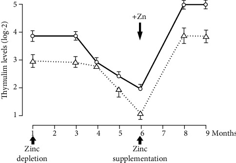
Zinc is an essential element for humans, and its deficiency was documented in 1963. Nutritional zinc deficiency is now known to affect over two billion subjects in the developing world. Conditioned deficiency of zinc in many diseases has also been observed. In zinc-deficient dwarfs from the Middle East, we reported growth retardation, delayed sexual development, susceptibility to infections, poor appetite, and mental lethargy. We never found a zinc-deficient dwarf who survived beyond the age of 25 y. In an experimental model of human mild zinc deficiency, we reported decreased thymulin (a thymopoietic hormone) activity in Th1 cells, decreased mRNAs of IL-2 and IFN-gamma genes, and decreased activity of natural killer cells (NK) and T cytotoxic T cells. The effect of zinc deficiency on thymulin activity and IL-2 mRNA was seen within eight to twelve weeks of the institution of zinc-deficient diet in human volunteers, whereas lymphocyte zinc decreased in 20 weeks and plasma zinc decreased in 24 weeks after instituting zinc-deficient diet. We hypothesized that decreased thymulin activity, which is known to proliferate Th1 cells, decreased the proliferation differentiation of Th1 cells. This resulted in decreased generation of IL-2 and IFN-gamma. We observed no effect in Th2 cell function; thus, zinc deficiency resulted in an imbalance of Th1 to Th2 function resulting in decreased cell-mediated immunity. Zinc therapy may be very useful in many chronic diseases. Zinc supplementation improves cell-mediated immunity, decreases oxidative stress, and decreases generation of chronic inflammatory cytokines in humans. Development of sensitive immunological biomarkers may be more sensitive than an assay of zinc in plasma and peripheral blood cells for diagnosis of marginal zinc deficiency in human.


Conflict of interest statement

The author declares no conflict of interest.

Figures

Figure 1
Figure 1
The landscape of zinc action on immune cells. Zinc is an essential component of thymulin, a thymic hormone involved in maturation and differentiation of T cells. The gene expression of IL-2 and IFN-γ (Th1 cytokines) is zinc dependent. IL-2 is involved in the activation of NK and T cytolytic cells. IL-12 is generated by stimulated macrophages-monocytes and is zinc dependent. IFN-γ and IL-12 together play a major role in the killing of parasites, viruses, and bacteria by macrophages-monocytes. Th2 cytokines are not affected by zinc deficiency except for IL-10 production, which is increased in the zinc-deficient elderly subjects. This is corrected by zinc supplementation. Increased IL-10 affects adversely Th1 and macrophage functions.
Figure 2
Figure 2
In a cell culture model HUT-78, the binding of NF-κB to DNA, an essential transcription factor for gene expression of Th1 cytokines (IL-2 and IFN-γ), is regulated by zinc as shown in this figure. We also show that the other transcription factors essential for gene expression of Th1 cytokines, AP1 and SP1, are also zinc dependent and their binding to DNA is regulated by zinc [35].
Figure 3
Figure 3
Our results of activation of NF-κB by zinc in HUT-78, a cell culture model. We observed that zinc was required for the expression of p105 mRNA, a precursor of p50 NF-κB protein. Once expressed, p50 NF-κB binds to IκB in the plasma. Following phosphorylation of IκB by zinc, NF-κB 50 is released for binding to DNA and gene expression of various proteins such as IL-2, IL-2Rα and β, IFN-γ, and IκB-α.
Figure 4
Figure 4
The translocation of NF-κB to DNA for binding and gene expression. Translocation of NF-κB-p50 to the nucleus under zinc-deficient and zinc-sufficient conditions. HUT-78 cells were incubated under Zn- and Zn+ conditions for 4 days and then exposed to PMA/PHA for 3 hours. Confocal images were prepared to show cytosolic NF-κB and nuclear material and the colocalization of NF-κB in the nucleus: (a) nonstimulated Zn- cells; (b) nonstimulated Zn+ cells; (c) PMA/PHA-stimulated Zn- cells; (d) PMA/PHA-stimulated Zn+ cells. Arrows indicate areas of colocalization. PMA/PHA-stimulated Zn+ cells showed the greatest translocation of NF-κB-p50 to the nucleus [36].
Figure 5
Figure 5
Activation of TCR increases intracellular free zinc. (a) Cells were incubated for 24 h in normal media to which had been added 15 μM zinc (to load cells with zinc) and then stimulated in normal media (5 μM zinc) in the presence of PHA, PMA, or Con-A for 30 and 60 min to determine which pathway was involved in differentiation or activation of Th0 or naïve cells and resulted in an increase in cellular free zinc. (b) To determine the time course for an increase in cellular free zinc, HUT-78 cells were stimulated in normal or in zinc-deficient medium. Stimulation in normal vs. zinc-deficient media allowed us to determine if the increase in cellular free zinc was due to an influx of extracellular free zinc or only a release of free zinc within the cellular compartment under Con-A stimulation. It appears that the increase in cellular free zinc is a result of both extracellular zinc influx and the release of free zinc from intracellular sources following Con-A stimulation (n = 3) [37].
Figure 6
Figure 6
Role of zinc in differentiating Th0 T cells to the Th1 subtype. The differentiation of Th0 or CD4+ naïve T cells to the Th1 subtype is a two-stage process involving many transcription factors and events which are zinc-dependent. (1) Engagement of TCR by CD3 antibodies or Con-A or an antigen by APC is the first step in the differentiation process. Here, zinc acts as a bridge between the CD4 or CD8 receptor and Lck, a tyrosine kinase activated during differentiation. (2) Once the initial process begins, zinc-dependent PKC-h is phosphorylated and able to activate the release of free zinc from metallothionein, the endoplasmic reticulum, or the Golgi. (3) An increase in free zinc is then used for binding of activated NF-κB-p50/p65 to DNA, which initiates the transcription and production of IFN-γ. (4) IFN-γ enhances the expression of T-bet. IFN-γ and T-bet expression then becomes autocrine/paracrine. During the second stage, after TCR is disengaged. (5) T-bet associates with STAT4 for transcription and expression of IL-12Rβ2 which is a zinc-dependent process. (6) Once the expression of IL-12Rβ2 is enhanced, T-bet expression then depends upon IL-12. (7) Once the Th1 cells are differentiated and stabilized, they function in the absence of IFN-γ. Full expression of T-bet also inhibits the expression of GATA3 which is responsible for Th2 differentiation, thus, locking in the Th1 phenotype [37].
Figure 7
Figure 7
Our concept regarding the role of zinc as an antioxidant and anti-inflammatory agent. Reactive oxygen species (ROS) is known to activate NF-κB. Zinc decreases ROS generation. NADPH oxidase is inhibited by zinc and SOD, which is both a zinc and copper-containing enzyme that is upregulated. SOD is known to decrease oxidative stress. Metallothionein (MT) is induced by zinc and MT, which contains 26 moles of cysteine per mole of protein, decreasing OH burden. Zinc via A20 inhibits NF-κB activation, and this results in a decrease in generation of inflammatory cytokines and adhesion molecules. This figure also shows that zinc may have a preventive role in some cancers such as colon and prostate and in atherosclerosis inasmuch as chronic inflammation has been implicated in the development of these disorders [46, 47].
Figure 8
Figure 8
Changes in plasma zinc during early and late zinc deficiency periods in marginal deficiency of zinc in humans. Plasma zinc levels (mean ± SD) during baseline (B) vs. early zinc deficiency period (E) and late zinc deficiency period (L) were as follows: B vs. E, 116.20 ± 3.51 μg/dl vs. 109.10 ± 8.30 μg/dl, p = 0.27; B vs. L, 116.20 ± 3.51 μg/dl vs. 105 ± 11.38 μg/dl, p = 0.23; and E vs. L, 109.10 ± 8.30 μg/dl vs. 105.53 ± 11.38 μg/dl, p = 0.31. The values for plasma zinc in normal control subjects (mean ± SD) are also shown (107.26 ± 8.92 μg/dl) [48].
Figure 9
Figure 9
Changes in lymphocyte zinc level during early and late zinc deficiency periods in the experimental model of human zinc deficiency. Lymphocyte zinc levels ((mean ± SD) μg/1010 cells) during baseline (B) vs. early zinc deficiency period (E) and late zinc deficiency period (L) were as follows: B vs. E, 58.36 ± 1.64 μg/1010 cells vs. 55.29 ± 4.20 μg/1010 cells, p = 0.25; B vs. L, 58.36 ± 1.64 μg/1010 cells vs. 41.67 ± 8.26 μg/1010 cells, p = 0.04; E vs. L, 55.29 ± 4.20 μg/1010 cells vs. 41.67 ± 8.26 μg/1010 cells, p = 0.03. The lymphocyte zinc level (mean ± SD) for control subjects is also shown (56.56 ± 6.42 μg/1010 cells [48].
Figure 10
Figure 10
Changes in lymphocyte 5′NT activity during baseline, early zinc deficiency, and late zinc deficiency periods in the experimental model of human zinc deficiency IL 5′NT activity (mean ± SD) nmoles IMP converted/106 lymphocytes/hour) during baseline (B) vs. early deficiency period (E) and late deficiency period (L) were as follows: B vs. E, 31.13 ± 5.56 nmol IMP converted per 106 lymphocytes per hour vs. 21.95 ± 0.92 nmol IMP converted per 106 lymphocytes per hour, p = 0.06; B vs. L, 31.13 ± 5.56 nmol IMP converted per 106 lymphocytes per hour vs. 18.50 ± 1.58 nmol IMP converted per 106 lymphocytes per hour, p = 0.03; E vs. L, 21.95 ± 0.92 nmol IMP converted per 106 lymphocytes per hour vs. 18.50 ± 1.58 nmol IMP converted per 106 lymphocytes per hour, p = 0.009. The values for 5′NT in normal control subjects are also shown (29.5 ± 6.53 nmol IMP converted per 106 lymphocytes per hour) [48].
Figure 11
Figure 11
Thymulin activity—levels of thymulin activity in a sequential study of young human volunteers submitted to a zinc-restricted diet for 6 mo followed by zinc supplementation are shown here. Results are expressed as log-2 reciprocal titers (mean ± SEM). Each determination was performed in triplicate [18].

References

    1. Raulin J. Chemical studies on vegetation. Annals of Science Nature. 1869;XI:93–99.
    1. Sommer A. L., Lipman C. B. Evidence on the indispensable nature of zinc and boron for higher green plants. Plant Physiology. 1926;1(3):231–249. doi: 10.1104/pp.1.3.231. - DOI - PMC - PubMed
    1. Todd W. R., Elvehjem C. A., Hart E. B. Zinc in the nutrition of the rat. American Journal of Physiology. 1933;107(1):146–156. doi: 10.1152/ajplegacy.1933.107.1.146. - DOI
    1. Tucker H. F., Salmon W. D. Parakeratosis or zinc deficiency disease in the Pig. Proceedings of the Society for Experimental Biology and Medicine. 1955;88(4):613–616. doi: 10.3181/00379727-88-21670. - DOI - PubMed
    1. O'Dell B. L., Newberne P. M., Savage J. E. Significance of dietary zinc for the growing chicken. Journal of Nutrition. 1958;65(4):503–523. doi: 10.1093/jn/65.4.503. - DOI - PubMed

MeSH terms