. 2020 Jul 10;6(7):1882-1893.
doi: 10.1021/acsinfecdis.0c00121. Epub 2020 May 29.

Potency Increase of Spiroketal Analogs of Membrane Inserting Indolyl Mannich Base Antimycobacterials Is Due to Acquisition of MmpL3 Inhibition

Affiliations

Potency Increase of Spiroketal Analogs of Membrane Inserting Indolyl Mannich Base Antimycobacterials Is Due to Acquisition of MmpL3 Inhibition

Ming Li et al. ACS Infect Dis. .

Abstract

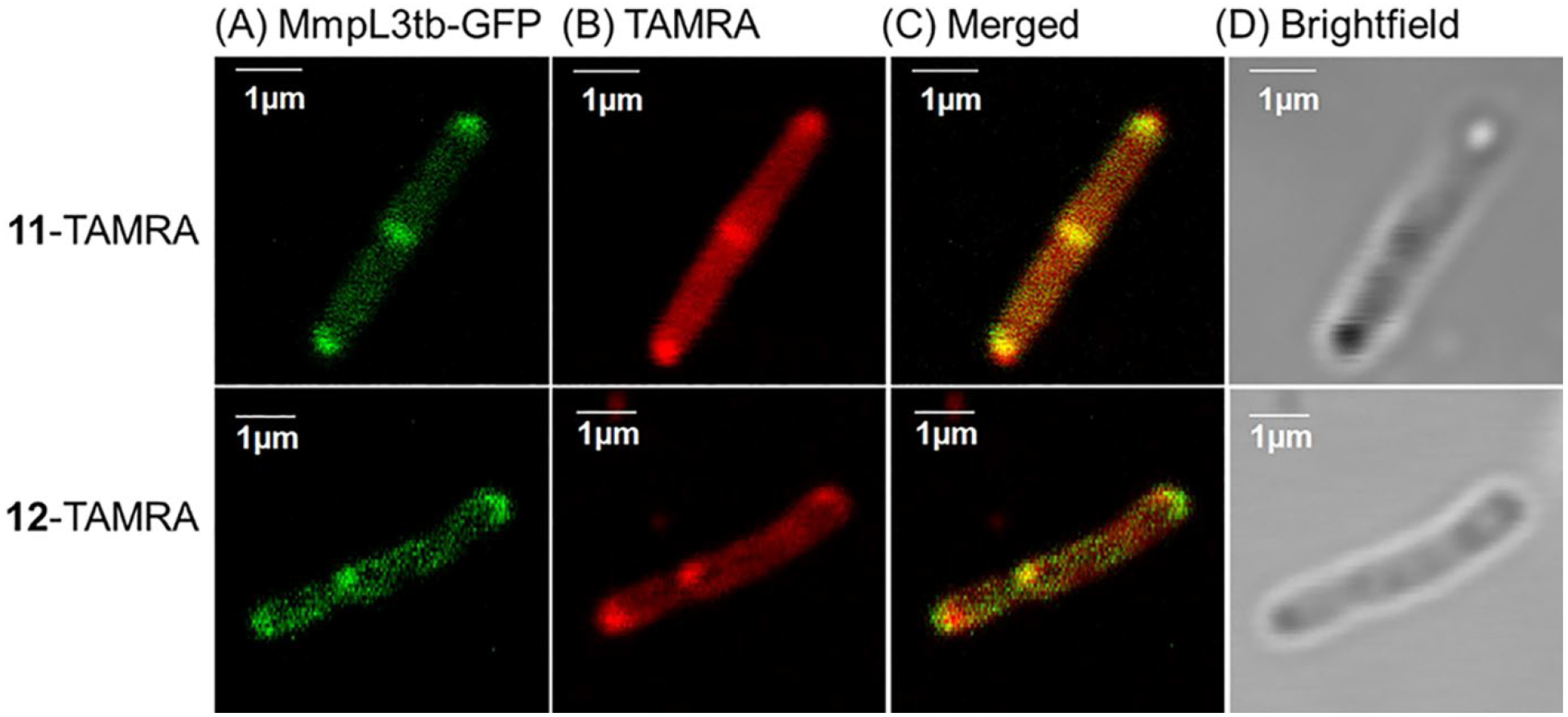
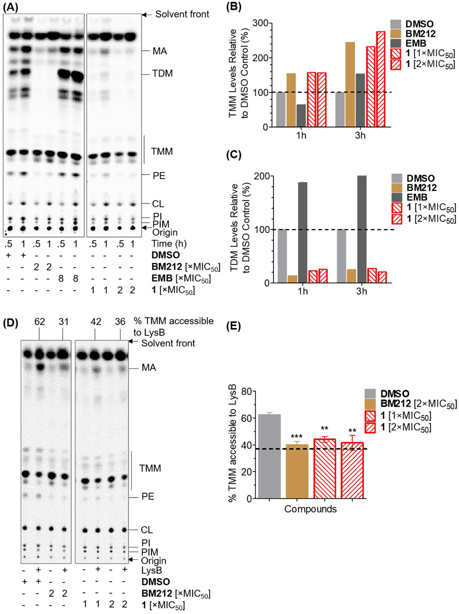
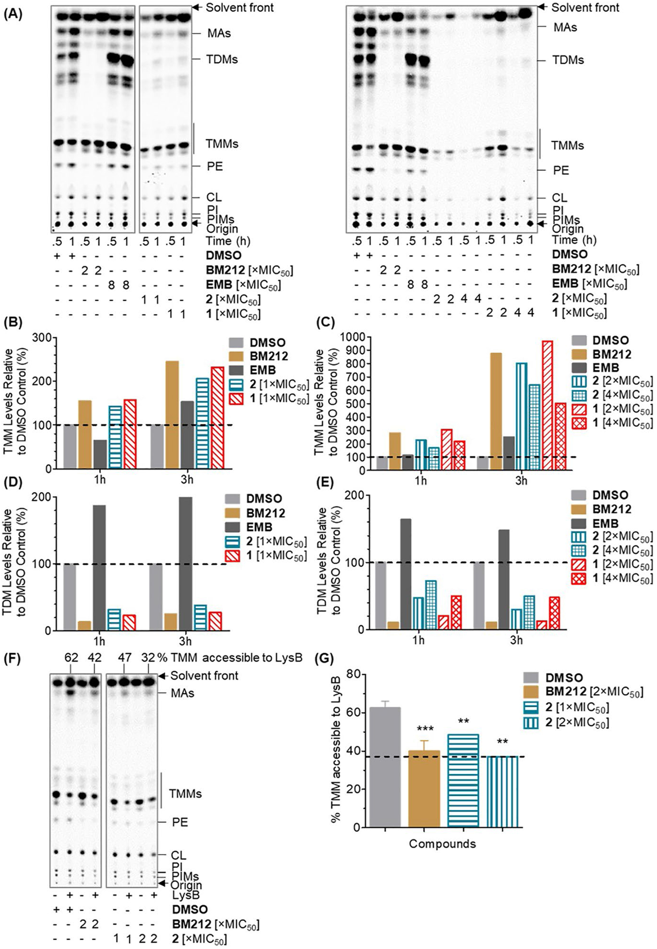
Chemistry campaigns identified amphiphilic indolyl Mannich bases as novel membrane-permeabilizing antimycobacterials. Spiroketal analogs of this series showed increased potency, and the lead compound 1 displayed efficacy in a mouse model of tuberculosis. Yet the mechanism by which the spiroketal moiety accomplished the potency "jump" remained unknown. Consistent with its membrane-permeabilizing mechanism, no resistant mutants could be isolated against indolyl Mannich base 2 lacking the spiroketal moiety. In contrast, mutations resistant against spiroketal analog 1 were obtained in mycobacterial membrane protein large 3 (MmpL3), a proton motive force (PMF)-dependent mycolate transporter. Thus, we hypothesized that the potency jump observed for 1 may be due to MmpL3 inhibition acquired by the addition of the spiroketal moiety. Here we showed that 1 inhibited MmpL3 flippase activity without loss of the PMF, colocalized with MmpL3tb-GFP in intact organisms, and yielded a consistent docking pose within the "common inhibitor binding pocket" of MmpL3. The presence of the spiroketal motif in 1 ostensibly augmented its interaction with MmpL3, an outcome not observed in the nonspiroketal analog 2, which displayed no cross-resistance to mmpL3 mutants, dissipated the PMF, and docked poorly in the MmpL3 binding pocket. Surprisingly, 2 inhibited MmpL3 flippase activity, which may be an epiphenomenon arising from its wider membrane disruptive effects. Hence, we conclude that the potency increase associated with the spiroketal analog 1 is linked to the acquisition of a second mechanism, MmpL3 inhibition. In contrast, the nonspiroketal analog 2 acts pleiotropically, affecting several cell membrane-embedded targets, including MmpL3, through its membrane permeabilizing and depolarizing effects.

Keywords: MmpL3; Mycobacterium tuberculosis; cationic amphiphiles; cell membrane; indolyl Mannich bases; membrane permeabilization.


Conflict of interest statement

The authors declare no competing financial interest.

Figures

Figure 1.
Figure 1.
Structures, antimycobacterial and selective cytotoxicity profiles of indolyl Mannich bases 1 and 2., The protonated Mannich base (blue) is an azaspiroketal in 1 and azepanyl in 2. Both have the lipophilic n-octyl side chain (red). clogP values are from ChemDraw Professional Version 15. 1.0.144.
Figure 2.
Figure 2.
Structures of azides (A) 11 and (B) 12. MIC50s of 11 and 12 were no more than 3× higher than those of 1 and 2, respectively.
Figure 3.
Figure 3.
Confocal microscopy images of MsmgΔmmpL3/pMVGH1-mmpL3tb-gfp cells treated with 11-TAMRA or 12-TAMRA conjugates. (A) GFP fluorescence of the MmpL3tb-GFP fusion protein at poles and septa. (B) Cells treated with 11-TAMRA or 12-TAMRA emitted red fluorescence with strong emissions from poles and septa. Unconjugated TAMRA-DBCO was not fluorescent (Figure S1). (C) Merged images from (A) and (B) confirmed overlap of green/red fluorescence at poles and septa. (D) Images under bright field. The images were acquired after sequential exposure to 11 (1×MIC50, 10 μM) or 12 (1×MIC50, 8 μM) for 3h, followed by TAMRA-DBCO (2h). Scale bar: 1 μm
Figure 4.
Figure 4.
(A) Predicted docking pose of 1 in the common inhibitor binding pocket of Msmg MmpL3 (PDB 6AJJ) showing H bonding between the protonated Mannich base (NH+) and D645, with NH⋯O distance of 1.99 Å. (B) 1 occupies subsites S3, S4 and S5 in the common inhibitor binding pocket of MmpL3. To note that the n-octyl side chain of 1 extends towards an open area beyond the binding channel in this pose. (C) Mapping of the resistance mutations of 1 on Msmg MmpL3 (PDB 6AJJ). S293 (red) is located within the common inhibitor binding pocket of MmpL3 (blue). (D) Location of the mutated residues S293 (TM-5), S596 (TM-9) and L325 (TM-6) in relation to 1 (in blue) in the binding pocket. (E) Representative consensus poses of 2 in Msmg MmpL3 (PDB 6AJJ) from Groups 1, 2 and 3. Asp-Tyr H-bonding was disrupted in Group 1 only. In Group 3, the n-octyl side chain occupied the entire length of S3 and S4, whereas in Groups 1 and 2, it projected out of the channel.
Figure 5.
Figure 5.
(A) A representative TLC of [14C]-labelled lipids extracted from Msmg. Cultures with DMSO represented non-drug treated negative control. Test compounds were MmpL3 inhibitor BM212 (31 μM, 2×MIC50), negative control ethambutol (EMB) (9.6 μM, 8×MIC50) and 1 (10 μM/20 μM, 1×/2×MIC50). (B) Quantification of TMM levels at indicated time points. TMM levels were quantified by densitometry from the TLC analysis in (A). Fraction of TMM in total mycolates (MA+TMM+TDM) in control and drug-treated samples were determined. For each drug-treated sample, the TMM/total mycolates ratio was normalized against the TMM/total mycolates ratio of DMSO control to give %TMM levels relative to DMSO control. (C) Quantification of TDM levels at indicated time points as described in (B). (D) A representative TLC of [14C]-labelled lipids extracted from Msmg spheroplasts treated with 1 or BM212 with or without LysB. (E) Graphical plot depicting the effects of 1 and BM212 on %TMM accessible to LysB in spheroplasts. To obtain %TMM accessible to LysB, fraction of TMM in total mycolates (MA+TMM) was determined for each sample. For each concentration of test compound, the difference in fraction of TMM levels between samples with and without LysB treatment was obtained and the value normalized against fraction of TMM in control non-LysB-treated spheroplasts. The dotted line is the %TMM accessible to LysB arising from random lysis of spheroplasts. This was 37% as described in Figure S5. Mean and SD from n=3 determinations. p < 0.01 (**) and < 0.001 (***) compared to DMSO control, Student’s t test, GraphPad Prism, Ver 5.0. Lipids monitored on the chromatograms were TMM, TDM, mycolic acid (MA), phosphatidylethanolamine (PE), cardiolipin (CL), phosphatidylinositol (PI) and phosphatidylinositol mannoside (PIM).
Figure 6.
Figure 6.
(A) Representative TLCs of [14C]-labelled lipids extracted from Msmg cells treated with BM212, ethambutol (EMB), 1 or 2. 2 was tested at 8 μM, 16 μM and 32 μM corresponding to 1×, 2× and 4×MIC50. BM212, EMB and 1 were tested at concentrations stated in Figure 5A. (B) and (C): Quantification of TMM levels at indicated time points. Lipid levels were quantified by densitometry and expressed in the same way as in Figure 5B. (D) and (E): Quantification of TDM levels at indicated time points. Lipid levels were quantified by densitometry and expressed in the same way as in Figure 5C. (F) A representative TLC of [14C]-labelled lipids extracted from compound-treated Msmg spheroplasts with or without LysB treatment. (G) Graphical plot depicting the effects of 2 and BM212 on % TMM accessible to LysB in spheroplasts. % TMM accessible to LysB was determined as described in Figure 5E. Mean and SD for n=3 determinations. p < 0.01 (**) and < 0.001 (***) compared to the DMSO control, Student’s t test, GraphPad Prism, Ver 5.0. Lipids monitored on the chromatograms were similar to those described in Figure 5.
Figure 7.
Figure 7.
Changes in membrane potential (ΔΨ) of Msmg spheroplasts treated with 1 or 2 at 2× and 4×MIC50. The strong increase in fluorescence signal (RFU) on addition of valinomycin+KCl at the 500s time point confirmed viability of spheroplasts for the duration of the experiment.

References

    1. The top 10 causes of death, https://www.who.int/news-room/fact-sheets/detail/thetop-10-causes-of-death (accessed March 11, 2020).
    1. World Health Organization (2019). Global tuberculosis report 2019. WHO Press, Geneva, Switzerland.
    1. FDA Approves New Treatment for Highly Drug-Resistant Forms of Tuberculosis, https://www.tballiance.org/news/fda-approves-new-treatment-highly-drug-r... (accessed March 11, 2020).
    1. Yang T, Moreira W, Nyantakyi SA, Chen H, Aziz DB, Go M-L, Dick T (2017) Amphiphilic indole derivatives as antimycobacterial agents: structure-activity relationships and membrane targeting properties. J. Med. Chem 60, 2745–2763. - PubMed
    1. Nyantakyi SA, Li M, Gopal P, Zimmerman M, Dartois V, Gengenbacher M, Dick T, Go M-L (2018) Indolyl azaspiroketal Mannich bases are potent antimycobacterial agents with selective membrane permeabilizing effects and in vivo activity. J. Med. Chem 61, 5733–5750. - PMC - PubMed

Publication types

LinkOut - more resources