. 2020 Jun 4;106(6):793-804.
doi: 10.1016/j.ajhg.2020.04.011. Epub 2020 May 14.

Expansion of GGC Repeat in GIPC1 Is Associated with Oculopharyngodistal Myopathy

Affiliations

Expansion of GGC Repeat in GIPC1 Is Associated with Oculopharyngodistal Myopathy

Jianwen Deng et al. Am J Hum Genet. .

Abstract

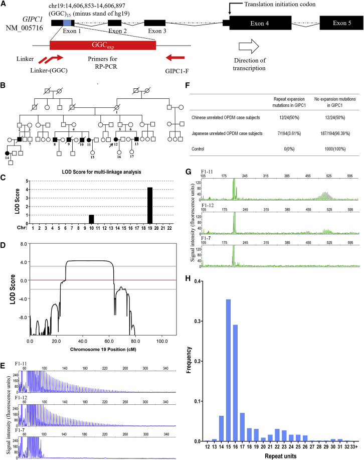
Oculopharyngodistal myopathy (OPDM) is an adult-onset inherited neuromuscular disorder characterized by progressive ptosis, external ophthalmoplegia, and weakness of the masseter, facial, pharyngeal, and distal limb muscles. The myopathological features are presence of rimmed vacuoles (RVs) in the muscle fibers and myopathic changes of differing severity. Inheritance is variable, with either putative autosomal-dominant or autosomal-recessive pattern. Here, using a comprehensive strategy combining whole-genome sequencing (WGS), long-read whole-genome sequencing (LRS), linkage analysis, repeat-primed polymerase chain reaction (RP-PCR), and fluorescence amplicon length analysis polymerase chain reaction (AL-PCR), we identified an abnormal GGC repeat expansion in the 5' UTR of GIPC1 in one out of four families and three sporadic case subjects from a Chinese OPDM cohort. Expanded GGC repeats were further confirmed as the cause of OPDM in an additional 2 out of 4 families and 6 out of 13 sporadic Chinese individuals with OPDM, as well as 7 out of 194 unrelated Japanese individuals with OPDM. Methylation, qRT-PCR, and western blot analysis indicated that GIPC1 mRNA levels were increased while protein levels were unaltered in OPDM-affected individuals. RNA sequencing indicated p53 signaling, vascular smooth muscle contraction, ubiquitin-mediated proteolysis, and ribosome pathways were involved in the pathogenic mechanisms of OPDM-affected individuals with GGC repeat expansion in GIPC1. This study provides further evidence that OPDM is associated with GGC repeat expansions in distinct genes and highly suggests that expanded GGC repeat units are essential in the pathogenesis of OPDM, regardless of the genes in which the expanded repeats are located.

Keywords: GGC repeat expansions; GIPC1; RNA-seq; intranuclear inclusions; oculopharyngodistal myopathy.


Conflict of interest statement

The authors declare no competing interests.

Figures

Figure 1
Figure 1
Detection of Expanded Repeats in the Long Reads Based on Comparisons between Healthy and Affected Individuals The flow chart shows the schematic of the discovery pipeline for repeat expansion. Healthy individuals (green boxes) and affected individuals (red boxes) with OPDM were sequenced using ONT PromethION. The same pipeline is used to implement the alignment and STR detection, and then construct the ERC matrix. A method, STR-Scoring, was developed to prioritize the STR.
Figure 2
Figure 2
Validation of GGC Repeat Expansions in GIPC1 and Variation of GGC Repeat Size among Normal Control Subjects (A) Schematic representation of GIPC1 indicating the location of GGC repeat expansions. The primer set used for RP-PCR analysis is designed to detect expanded GGC repeats (red line and arrows). (B) Pedigree chart of a family with OPDM (family 1). Squares and circles indicate males and females, respectively. A diagonal line through a symbol indicates a deceased individual. Affected individuals are indicated by filled symbols. The pedigree charts are simplified for confidentiality reasons. All the numbered individuals in family 1 were examined by RP-PCR and AL-PCR. As shown in the pedigree chart, seven individuals had repeat expansion variations, whereas ten unaffected individuals, including three married-in individuals, did not have repeat expansion mutations. (C) Multipoint parametric linkage analysis demonstrated a linkage interval with maximum logarithm of the odds (LOD) scores of 4.21 in chromosome 19. (D) Genetic linkage analysis indicated maximum LOD scores of 4.21 in chromosome 19, a 29.18-Mb region at 19p13.2–19q13.12 (chr19: 8,841,079–38,022,358). (E) Representative results of RP-PCR analysis showing GGC repeat expansions of individuals in family 1 (top and middle panels). In an unaffected individual from family 1, no GGC repeat expansions were detected (bottom panel). Experiments were conducted thrice with reproducible results. (F) GGC repeat expansions in GIPC1 were observed in 12 out of the 24 Chinese OPDM-affected case subjects (8 familial and 16 sporadic case subjects). The repeat expansion mutations were also detected in 7 out of 194 Japanese individuals, but were not detected in any of the 1,000 control subjects. (G) Representative results of AL-PCR analysis showing the numbers of expanded GGC repeats of individuals in family 1 (top and middle panels). In an unaffected individual from family 1, no GGC repeat expansions were detected (bottom panel). (H) Frequency distribution of repeat units of GGC repeats of 550 control subjects in GIPC1, as revealed by fragment analysis.
Figure 3
Figure 3
Muscle MRI and Myopathological Changes of the GIPC1-Affected Individuals with OPDM (A and B) Muscle MRI of individual S3 showed fatty infiltration of lower limb muscles, with the distal muscles (B, calf level) more severely affected than the proximal muscles (A, thigh level). (C–F) Hematoxylin & eosin (H&E) (C and D) and modified Gomori trichrome (mGT) (E and F) staining of muscle sections from individuals S6 and S7, showing dystrophic change with variation in fiber size and endomysial fibrosis, and fibers with rimmed vacuoles (marked by arrow). (G and H) P62 staining of muscle section from individual S6, inclusion bodies shown in both rimmed vacuole (RV) (marked by arrow) and nuclei (marked by arrowhead and higher magnification). (I) Electron microscopy of muscle tissue from individual S7. RV filled with myeloid body and higher magnification showed cytoplasmic tubulofilamentous inclusion bodies (marked by arrow). (J) Electron microscopy of muscle tissue from individual F1-12. Intranuclear inclusions contained filamentous aggregates in the nuclei of the muscle fiber (marked by arrowhead and shown at higher magnification).
Figure 4
Figure 4
Methylation and Expression at GIPC1 Locus (A) Methylation status across the expanded GGC repeat region in GIPC1 from whole blood DNA was determined using LRS data from five affected individuals (F1-11, F1-12, S2, S3, S4) and 100 healthy individuals; no significant difference in methylation was detected between OPDM-affected individuals and control subjects. (B) Quantification of methylation level between OPDM-affected individuals and control subjects. (C) Methylation status between expanded and non-expanded alleles of GIPC1 from whole blood DNA was determined using LRS data from five affected individuals (F1-11, F1-12, S2, S3, S4), and no significant difference was detected between expanded and non-expanded alleles. (D) Quantification of methylation level between expanded and non-expanded alleles. All data were analyzed using Wilcoxon rank sum test in (A) and (C), or Student’s t test in (B) and (D). (E) Western blot analysis of GIPC1 protein levels in five OPDM-affected individuals with expanded GGC repeats in GIPC1 and four age-matched control subjects. GAPDH was used as a loading control. (F) Quantification of relative GIPC1 protein level in each group; no significant difference was observed between OPDM-affected individuals with expanded GGC repeats and controls. Data were analyzed using Student’s t test; ns, not significant.
Figure 5
Figure 5
GIPC1 Distribution in Skeletal Muscle and RNA-Seq Profiling (A) Immunofluorescence staining reveals the distribution of GIPC1 in muscle fibers. GIPC1 partially co-localized with: p62-positive inclusions in rimmed vacuole marked with arrows; and p62-positive intranuclear inclusions marked with arrowheads. (B) Heatmap showing hierarchical clustering of the differentially expressed mRNAs, with annotated genes in the p53 signaling pathway, vascular smooth muscle contraction, ubiquitin-mediated proteolysis, and ribosome.

References

    1. Durmus H., Laval S.H., Deymeer F., Parman Y., Kiyan E., Gokyigiti M., Ertekin C., Ercan I., Solakoglu S., Karcagi V. Oculopharyngodistal myopathy is a distinct entity: clinical and genetic features of 47 patients. Neurology. 2011;76:227–235. - PubMed
    1. Satoyoshi E., Kinoshita M. Oculopharyngodistal myopathy. Arch. Neurol. 1977;34:89–92. - PubMed
    1. Lu H., Luan X., Yuan Y., Dong M., Sun W., Yan C. The clinical and myopathological features of oculopharyngodistal myopathy in a Chinese family. Neuropathology. 2008;28:599–603. - PubMed
    1. Minami N., Ikezoe K., Kuroda H., Nakabayashi H., Satoyoshi E., Nonaka I. Oculopharyngodistal myopathy is genetically heterogeneous and most cases are distinct from oculopharyngeal muscular dystrophy. Neuromuscul. Disord. 2001;11:699–702. - PubMed
    1. Mignarri A., Carluccio M.A., Malandrini A., Sicurelli F., Galli L., Mazzei M.A., Federico A., Orrico A., Dotti M.T. The first Italian patient with oculopharyngodistal myopathy: case report and considerations on differential diagnosis. Neuromuscul. Disord. 2012;22:759–762. - PubMed

Publication types

MeSH terms

Substances

Supplementary concepts