. 2020 May 13;9(5):417.
doi: 10.3390/antiox9050417.

Palmitic Acid Affects Intestinal Epithelial Barrier Integrity and Permeability In Vitro

Affiliations

Palmitic Acid Affects Intestinal Epithelial Barrier Integrity and Permeability In Vitro

Manuele Gori et al. Antioxidants (Basel). .

Abstract

Palmitic acid (PA), a long-chain saturated fatty acid, might activate innate immune cells. PA plays a role in chronic liver disease, diabetes and Crohn's disease, all of which are associated with impaired intestinal permeability. We investigated the effect of PA, at physiological postprandial intestinal concentrations, on gut epithelium as compared to lipopolysaccharide (LPS) and ethanol, using an in vitro gut model, the human intestinal epithelial cell line Caco-2 grown on transwell inserts. Cytotoxicity and oxidative stress were evaluated; epithelial barrier integrity was investigated by measuring the paracellular flux of fluorescein, and through RT-qPCR and immunofluorescence of tight junction (TJ) and adherens junction (AJ) mRNAs and proteins, respectively. In PA-exposed Caco-2 monolayers, cytotoxicity and oxidative stress were not detected. A significant increase in fluorescein flux was observed in PA-treated monolayers, after 90 min and up to 360 min, whereas with LPS and ethanol, this was only observed at later time-points. Gene expression and immunofluorescence analysis showed TJ and AJ alterations only in PA-exposed monolayers. In conclusion, PA affected intestinal permeability without inducing cytotoxicity or oxidative stress. This effect seemed to be faster and stronger than those with LPS and ethanol. Thus, we hypothesized that PA, besides having an immunomodulatory effect, might play a role in inflammatory and functional intestinal disorders in which the intestinal permeability is altered.

Keywords: Caco-2 monolayer; adherens junctions.; gut barrier integrity; intestinal permeability; palmitic acid; tight junctions.


Conflict of interest statement

The authors declare no conflict of interest.

Figures

Figure 1
Figure 1
Analysis of cell viability/cytotoxicity following the treatments of Caco-2 monolayers with lipopolysaccharide (LPS), ethanol, and palmitic acid (PA). (a) LPS (10 μg/mL) for 24 h. (b) Ethanol (10%) for 1 h. (c) PA (1 mM) for 24 h. The histograms show the percentage of living cells for the control (dashed lines, referred to as 100%), treated (grey bars), and positive control cells (black bars, by using 2% Triton X-100 for 2 h) of human colonic mucosa. Representative micrographs of the different treatments show the nuclei of dead cells (green), through the Blue/Green Cell Viability Imaging Kit, for both the treated (lower panels in ad) and the control cells (middle panels in ad). Each treatment was compared to its own internal control. Data are reported as mean ± SEM; n = 3 independent experiments. Statistical analysis was performed using the unpaired t-test. **** p < 0.0001. Scale bars: 50 μm. Ctrl (Control); LPS (Lipopolysaccharide); EtOH (Ethanol); PA (Palmitic Acid); and Pos. Ctrl (Positive Control).
Figure 2
Figure 2
Reactive oxygen species (ROS) production for the analysis of oxidative stress following the treatments of Caco-2 monolayers with LPS, ethanol, and PA. The histograms show the fold increase of MFI expressed as the ratio between the treated (grey bars) and control (dashed lines, referred to as 1) cells, for LPS (a), ethanol (b), and PA (c); positive control cells (black bars in ac) were exposed to 400 μM H2O2 for 3 h. Representative intensity surface plots of the control and treated cells for LPS (in a, middle and lower panels, respectively), ethanol (in b, middle and lower panels, respectively), PA (in c, middle and lower panels, respectively), and positive control (in d, middle and lower panels, respectively). Data are reported as mean ± SEM; n = 3 independent experiments. Statistical analysis was performed using unpaired t-test. *** p < 0.001. Ctrl (Control); LPS (Lipopolysaccharide); EtOH (Ethanol); PA (Palmitic Acid); and Pos. Ctrl (Positive Control).
Figure 3
Figure 3
Barrier function of the Caco-2 cell monolayers analyzed through the FD-4 permeability assay. (a) LPS treatment (10 μg/mL) for 24 h. (b) Ethanol treatment (10%) for 1 h. (c) PA treatment (1 mM) for 24 h and washout (W), with a 24 h recovery after PA treatment (1 mM) for 24 h. The amount of FD-4 (expressed in pmol) accumulated in the receiver compartment, was plotted as a function of time (expressed in min)—in red for each treatment (square dots and triangular dots) and in black for the internal control (round dots)—with independent controls among the three conditions. Data are reported as mean ± SEM; n = 4 independent experiments. Statistical analysis was performed using a two-way ANOVA, followed by Bonferroni’s post-hoc correction test. * p < 0.05, ** p < 0.01, *** p < 0.001, **** p < 0.0001. Ctrl (Control); LPS (Lipopolysaccharide); EtOH (Ethanol); PA (Palmitic Acid); and W (Washout).
Figure 4
Figure 4
Gene expression analysis of the intestinal barrier complexes. Gene expression of tight junction protein 1 (TJP1), E-cadherin (CDH1), and β-catenin (CTNNB1) in the Caco-2 monolayers treated with LPS for 24h (ac), with ethanol for 1 h (df), and with PA for 24 h (gi), compared to the internal controls. Data are presented as relative expression of mRNAs vs. controls and reported as mean ± SEM; n = 3 independent experiments. Statistical analysis was performed using the unpaired t-test. ** p < 0.01 and *** p < 0.001. Ctrl (Control); LPS (Lipopolysaccharide); EtOH (Ethanol); and PA (Palmitic Acid).
Figure 5
Figure 5
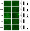
Immunohistochemical analysis of the intestinal barrier proteins. Differential protein expression of E-cadherin (CDH1) and β-catenin (CTNNB1) between the control (a,b, far left panels) and ethanol-treated (a,b, middle panels) Caco2 cells, with a discontinuous expression on the cell surface, as shown in the representative micrographs; in (a, far right panel) and (b, far right panel) the histograms show the relative expression of the fluorescence intensity (FITC) on the cell surface of the Caco-2 monolayers treated with ethanol vs. the controls, through perimeter analysis. Differential expression of tight junction protein 1 (TJP1), CDH1, and CTNNB1 between the control (c,d,e, far left panels, respectively) and the PA-treated (c,d,e, middle panels, respectively) Caco2 cells; in (c), (d), and (e) the far right histograms show the relative expression of the fluorescence intensity (FITC) on the cell surface of Caco-2 monolayers treated with PA vs. the controls, as for the far right histograms of (a) and (b). The immunostainings against CDH1 in the control (d, far left panel) and PA-treated (d, middle panel) Caco-2 cells also highlight the different morphology and size of the PA-treated vs. control cells. Data are reported as mean ± SEM; n = 3 independent experiments. Statistical analysis was performed using unpaired t-test. * p < 0.05, ** p < 0.01, *** p < 0.001. Scale bars: 25 μm. Ctrl (Control); EtOH (Ethanol); and PA (Palmitic Acid).

References

    1. Camilleri M., Madsen K., Spiller R.C., Meerveld B.G.-V., Verne G.N. Intestinal barrier function in health and gastrointestinal disease. Neurogastroenterol. Motil. 2012;24:503–512. doi: 10.1111/j.1365-2982.2012.01921.x. - DOI - PMC - PubMed
    1. Hering N.A., Fromm M., Schulzke J.-D. Determinants of colonic barrier function in inflammatory bowel disease and potential therapeutics. J. Physiol. 2012;590:1035–1044. doi: 10.1113/jphysiol.2011.224568. - DOI - PMC - PubMed
    1. Kolosov D., Chasiotis H., Kelly S., Stellwagen S.D., Opell B.D., Short K.G. Tight junction protein gene expression patterns and changes in transcript abundance during development of model fish gill epithelia. J. Exp. Boil. 2014;217:1667–1681. doi: 10.1242/jeb.098731. - DOI - PubMed
    1. Bein A., Zilbershtein A., Golosovsky M., Davidov D., Schwartz B. LPS Induces Hyper-Permeability of Intestinal Epithelial Cells. J. Cell. Physiol. 2016;232:381–390. doi: 10.1002/jcp.25435. - DOI - PubMed
    1. Guarino M.P.L., Sessa R., Altomare A., Cocca S., Di Pietro M., Carotti S., Schiavoni G., Alloni R., Emerenziani S., Morini S., et al. Human colonic myogenic dysfunction induced by mucosal lipopolysaccharide translocation and oxidative stress. Dig. Liver Dis. 2013;45:1011–1016. doi: 10.1016/j.dld.2013.06.001. - DOI - PubMed

LinkOut - more resources