. 2020 May 13;12(5):1227.
doi: 10.3390/cancers12051227.

LINC00261 Is Differentially Expressed in Pancreatic Cancer Subtypes and Regulates a Pro-Epithelial Cell Identity

Affiliations

LINC00261 Is Differentially Expressed in Pancreatic Cancer Subtypes and Regulates a Pro-Epithelial Cell Identity

Agnes Dorn et al. Cancers (Basel). .

Abstract

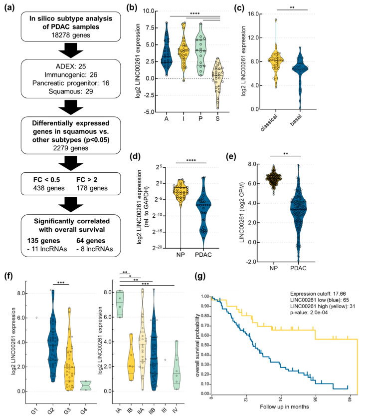
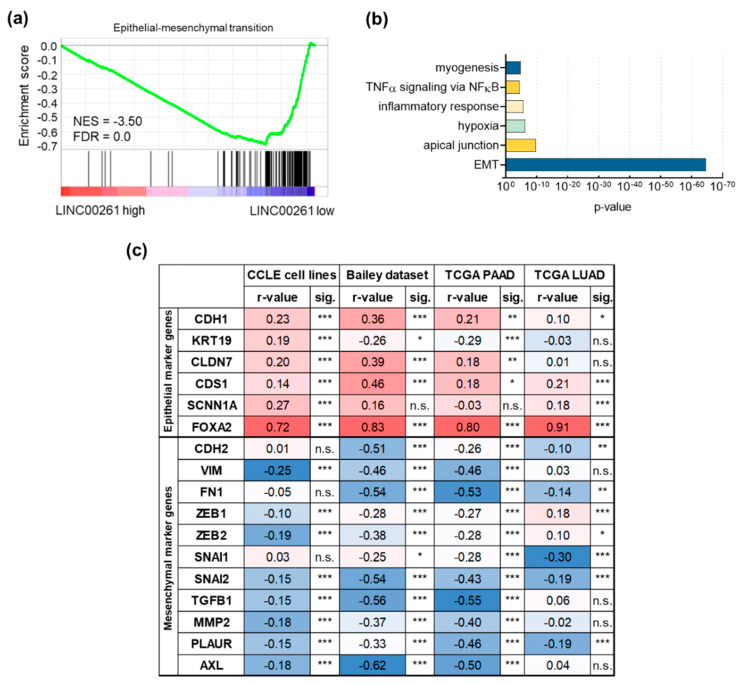
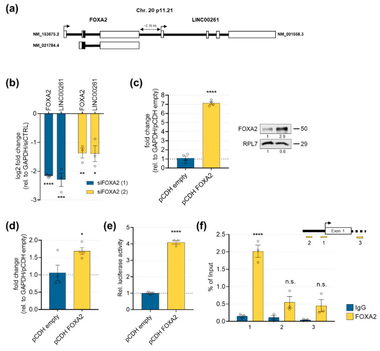
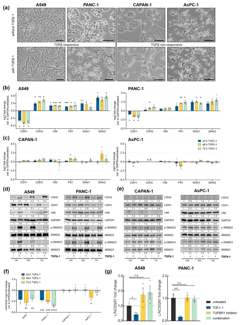
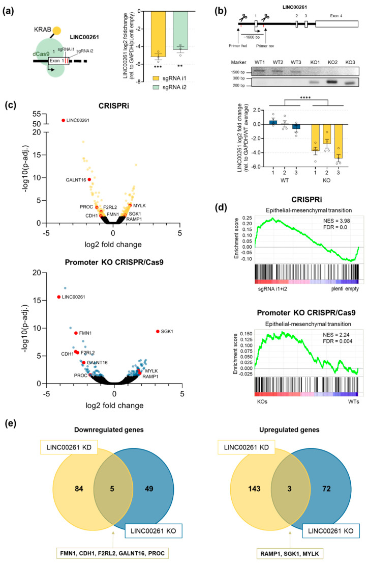
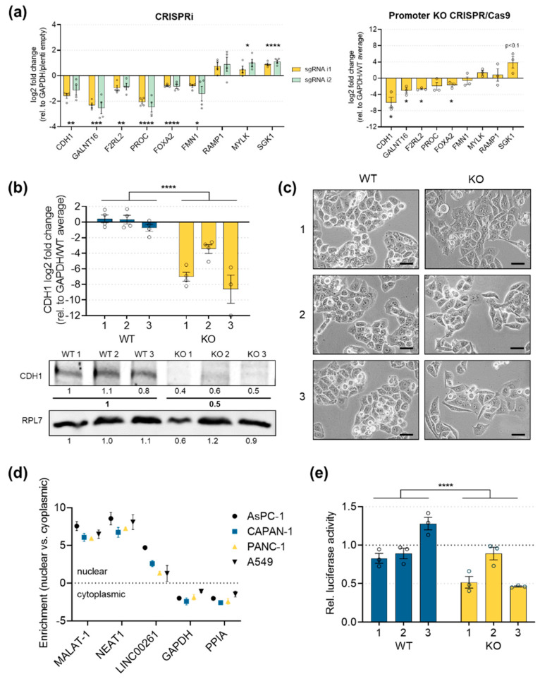
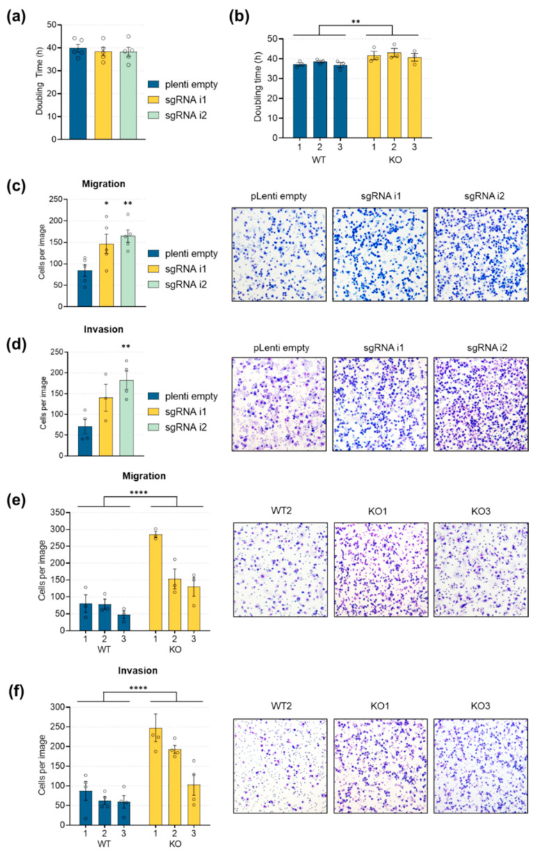
Pancreatic adenocarcinoma (PDAC) is one of the major causes of cancer-associated deaths worldwide, with a dismal prognosis that has not significantly changed over the last decades. Transcriptional analysis has provided valuable insights into pancreatic tumorigenesis. Specifically, pancreatic cancer subtypes were identified, characterized by specific mutations and gene expression changes associated with differences in patient survival. In addition to differentially regulated mRNAs, non-coding RNAs, including long non-coding RNAs (lncRNAs), were shown to have subtype-specific expression patterns. Hence, we aimed to characterize prognostic lncRNAs with deregulated expression in the squamous subtype of PDAC, which has the worst prognosis. Extensive in silico analyses followed by in vitro experiments identified long intergenic non-coding RNA 261 (LINC00261) as a downregulated lncRNA in the squamous subtype of PDAC, which is generally associated with transforming growth factor β (TGFβ) signaling in human cancer cells. Its genomic neighbor, the transcription factor forkhead box protein A2 (FOXA2), regulated LINC00261 expression by direct binding of the LINC00261 promoter. CRISPR-mediated knockdown and promoter knockout validated the importance of LINC00261 in TGFβ-mediated epithelial-mesenchymal transition (EMT) and established the epithelial marker E-cadherin, an important cell adhesion protein, as a downstream target of LINC00261. Consequently, depletion of LINC00261 enhanced motility and invasiveness of PANC-1 cells in vitro. Altogether, our data suggest that LINC00261 is an important tumor-suppressive lncRNA in PDAC that is involved in maintaining a pro-epithelial state associated with favorable disease outcome.

Keywords: CDH1; EMT; FOXA2; LINC00261; PDAC; TGFβ; lncRNA.


Conflict of interest statement

The authors declare no conflict of interest.

Figures

Figure 1
Figure 1
Analysis of long intergenic non-coding RNA 261 (LINC00261) expression in pancreatic adenocarcinoma (PDAC). (a) Flow chart that outlines the identification of candidate long non-coding RNAs (lncRNAs) important for PDAC progression and patient survival; ADEX, aberrantly differentiated endocrine exocrine; FC, fold change. (b) Analysis of the Bailey PDAC dataset revealed a significant downregulation of LINC00261 expression in the squamous (S) compared to the pancreatic progenitor (P), immunogenic (I), and ADEX (A) subtypes (**** p < 0.0001, one-way ANOVA). (c) Analysis of the pancreatic adenocarcinoma dataset from The Cancer Genome Atlas (TCGA) according to Moffitt’s classification highlighted significant downregulation of LINC00261 expression in the basal-like compared to the classical subtype (** p < 0.01, unpaired t-test). (d,e) Analysis of LINC00261 expression in 34 normal pancreatic (NP) tissues, 42 PDAC tissues (d), and in publicly available TCGA and Genotype-Tissue Expression (GTEx) datasets (e) (normal pancreas: n = 177, PDAC: n = 248) showed significantly lower LINC00261 expression in pancreas adenocarcinoma compared to normal pancreas (** p < 0.01, **** p < 0.0001, Mann–Whitney U test). (f) LINC00261 expression is significantly lower in high grade (G1: n = 1, G2: n = 56, G3: n = 34, G4: n = 2) and high-stage tumors (IA: n = 4, IB: n = 5, IIA: n = 25, IIB: n = 55, III: n = 1, IV: n = 6); * p < 0.05, ** p < 0.01, *** p < 0.001, **** p < 0.0001, one-way ANOVA. (g) Survival analysis for PDAC patients with low LINC00261 (blue line, n = 65) versus high LINC00261 (yellow line, n = 31) expression (Bailey dataset, http://r2.amc.nl, Log rank test).
Figure 2
Figure 2
Association of long intergenic non-coding RNA 261 (LINC00261) with epithelial–mesenchymal transition (EMT). (a) Gene set enrichment analysis (GSEA) of PDAC samples with low (n = 48) versus high LINC00261 expression (n = 48). Median LINC00261 expression was used as a cut-off; NES, normalized enrichment score; FDR, false discovery rate. (b) GSEA analysis of genes that were significantly inversely correlated with LINC00261 in the pancreatic adenocarcinoma (PDAC) dataset of Bailey et al. identified EMT as the highest enriched hallmark gene set. (c) Correlation of selected epithelial and mesenchymal marker genes with LINC00261 expression (* p < 0.05, ** p < 0.01, *** p < 0.001). Note: CDH1, E-cadherin; KRT19, keratin 19; CLDN7, claudin 7; CDS1, CDP-diacylglycerol synthase 1; SCNN1A, sodium channel epithelial 1 subunit alpha; FOXA2, forkhead box A2; CDH2, N-cadherin; VIM, vimentin; FN1, fibronectin 1; ZEB1, zinc finger E-box binding homeobox 1; ZEB2, zinc finger E-box binding homeobox 2; SNAI1, snail family transcriptional repressor 1; SNAI2, snail family transcriptional repressor 2; TGFB1, transforming growth factor beta 1; MMP2, matrix metallopeptidase 2; PLAUR, plasminogen activator, urokinase receptor; AXL, AXL receptor tyrosine kinase.
Figure 3
Figure 3
LINC00261 regulation by its genomic neighbor FOXA2. (a) The genomic loci of LINC00261 and FOXA2 on chromosome 20. (b) The siRNA-mediated knockdown of FOXA2 significantly downregulated FOXA2 and LINC00261 RNA levels (* p < 0.05, ** p < 0.01, *** p < 0.001, **** p < 0.0001, one-way ANOVA). (c) Stable overexpression of FOXA2 in PANC-1 was achieved at RNA (left panel; glyceraldehyde-3-phosphate dehydrogenase (GAPDH) was used as the reference gene) and protein levels (right panel; ribosomal protein L7 (RPL7) was used as a loading control; **** p < 0.0001, unpaired t-test). (d) LINC00261 expression levels in control or FOXA2 overexpressing PANC-1 cells (* p < 0.05, unpaired t-test). (e) Luciferase activity of a LINC00261 promoter reporter after stable FOXA2 or empty control vector overexpression in PANC-1 cells (**** p < 0.0001, unpaired t-test). (f) Chromatin immunoprecipitation (ChIP) followed by qPCR analysis using primers located upstream (1,2) and downstream (3) of the LINC00261 transcriptional start site (upper panel) confirmed binding of FOXA2 to the LINC00261 promoter region (lower panel, **** p < 0.0001, unpaired t-test).
Figure 4
Figure 4
Transforming growth factor β (TGFβ) downregulates LINC00261 expression in cell lines that show TGFβ-induced EMT. (a) Brightfield images of untreated and TGFβ-1 treated A549, PANC-1 (TGFβ-responsive), AsPC-1, and CAPAN-1 cells (TGFβ-non-responsive). Scale bar = 100 µm. Analysis of mRNA (b,c) and protein levels (d,e) of genes associated with EMT was done by qRT-PCR and Western Blot, respectively (* p < 0.05, ** p < 0.01, *** p < 0.001, two-way ANOVA). (f) LINC00261 expression in TGFβ-responsive and non-responsive cells using qRT-PCR (* p < 0.05, ** p < 0.01, *** p < 0.001, **** p < 0.0001, unpaired t-test). (g) LINC00261 regulation in A549 and PANC-1 cells treated with TGFβ-1, TGFBR1 inhibitor (RepSox), or both after 72 h, measured by qRT-PCR (* p < 0.05, ** p < 0.01, unpaired t-test).
Figure 5
Figure 5
RNA-seq analysis revealed an enrichment of the EMT gene set in LINC00621low PANC-1 cells. (a) Schema of CRISPR interference (CRISPRi)-mediated targeting of LINC00261 and its expression levels in PANC-1 cells measured by qRT-PCR (** p < 0.01, *** p < 0.001, one-way ANOVA). (b) Schema of CRISPR/Cas9-mediated knockout of the promoter region of LINC00261 using two sgRNAs. Cutting of both sgRNAs led to the removal of a genomic fragment of ~1600 bp. PCR and gel electrophoresis using the indicated primers resulted in a ~1600 bp product in wild-type clones and a ~250 bp product in knockout clones (**** p < 0.0001, two-way ANOVA). (c) Volcano plots of RNA-seq analyses of differently expressed genes of CRISPRi- and CRISPR/Cas9-mediated knockdown of LINC00261. Significantly deregulated genes (adjusted p-value (p-adj.) < 0.05) are highlighted. Genes found to be deregulated in both cell systems are labelled in red. (d) GSEA analysis of RNA sequencing data revealed a significant enrichment of the EMT gene set in LINC00261-depleted cells in both knockdown systems. (e) Venn diagrams show the intersection of significantly (p-adj. < 0.05) downregulated or upregulated genes due to LINC00261 knockdown by CRISPRi or CRISPR/Cas9 systems.
Figure 6
Figure 6
LINC00261 regulated E-cadherin expression. (a) The qPCR analysis of the genes commonly deregulated in both CRISPR systems, as shown in Figure 5e, in addition to FOXA2 (* p < 0.05, ** p < 0.01, *** p < 0.001, **** p < 0.0001, two-way ANOVA, unpaired t-test). (b) Expression of E-cadherin mRNA (upper panel) and protein levels (lower panel) in three wild-type and three promoter knockout clones (**** p < 0.0001, two-way ANOVA). (c) Brightfield images of wild-type and LINC00261 promoter knockout clones (20× objective, scale bar = 50 µm). (d) Cellular fractionation in lung cancer and pancreatic cancer cell lines, highlighting nuclear enrichment of LINC00261. (e) Relative luciferase activity of the CDH1 gene promoter constructs normalized to the pGL3-vector with a minimal cytomegalovirus (CMV) promotor (pGL3-minCMV). The average of the relative luciferase activity in wild-type clones was set to 1.0 (**** p < 0.0001, two-way ANOVA).
Figure 7
Figure 7
Enhanced cell migration and invasion in LINC00261low cells. (a,b) Cell doubling time in PANC-1 cells after CRISPRi-mediated LINC00261 downregulation and in three wild-type and three LINC00261 promoter knockout cell clones (** p < 0.01, one-way (a) and two-way ANOVA (b)). (c,d) Transwell migration (c) and invasion (d) assays in PANC-1 cells after CRISPRi-mediated LINC00261 downregulation (* p < 0.05, ** p < 0.01, one-way ANOVA). (e,f) Transwell migration (e) and invasion (f) assays in PANC-1 cells after CRISPR/Cas9-mediated knockout of LINC00261 promoter. Quantification of migrated and invaded cells (cf) from five random fields of three wild-type and three knockout clones after eosin Y and methylene blue staining using light microscopy (**** p < 0.0001, two-way ANOVA).

References

    1. Siegel R.L., Miller K.D., Jemal A. Cancer Statistics, 2017. CA Cancer J. Clin. 2017;67:7–30. doi: 10.3322/caac.21387. - DOI - PubMed
    1. Rahib L., Smith B.D., Aizenberg R., Rosenzweig A.B., Fleshman J.M., Matrisian L.M. Projecting cancer incidence and deaths to 2030: The unexpected burden of thyroid, liver, and pancreas cancers in the United States. Cancer Res. 2014;74:2913–2921. doi: 10.1158/0008-5472.CAN-14-0155. - DOI - PubMed
    1. Bailey P., Chang D.K., Nones K., Johns A.L., Patch A.M., Gingras M.C., Miller D.K., Christ A.N., Bruxner T.J., Quinn M.C., et al. Genomic analyses identify molecular subtypes of pancreatic cancer. Nature. 2016;531:47–52. doi: 10.1038/nature16965. - DOI - PubMed
    1. Collisson E.A., Sadanandam A., Olson P., Gibb W.J., Truitt M., Gu S., Cooc J., Weinkle J., Kim G.E., Jakkula L., et al. Subtypes of pancreatic ductal adenocarcinoma and their differing responses to therapy. Nat. Med. 2011;17:500–503. doi: 10.1038/nm.2344. - DOI - PMC - PubMed
    1. Moffitt R.A., Marayati R., Flate E.L., Volmar K.E., Loeza S.G., Hoadley K.A., Rashid N.U., Williams L.A., Eaton S.C., Chung A.H., et al. Virtual microdissection identifies distinct tumor- and stroma-specific subtypes of pancreatic ductal adenocarcinoma. Nat. Genet. 2015;47:1168–1178. doi: 10.1038/ng.3398. - DOI - PMC - PubMed

LinkOut - more resources