. 2020 Jul 3;295(27):9223-9243.
doi: 10.1074/jbc.RA120.013792. Epub 2020 May 15.

A terminal α3-galactose modification regulates an E3 ubiquitin ligase subunit in Toxoplasma gondii

Affiliations

A terminal α3-galactose modification regulates an E3 ubiquitin ligase subunit in Toxoplasma gondii

Msano Mandalasi et al. J Biol Chem. .

Abstract

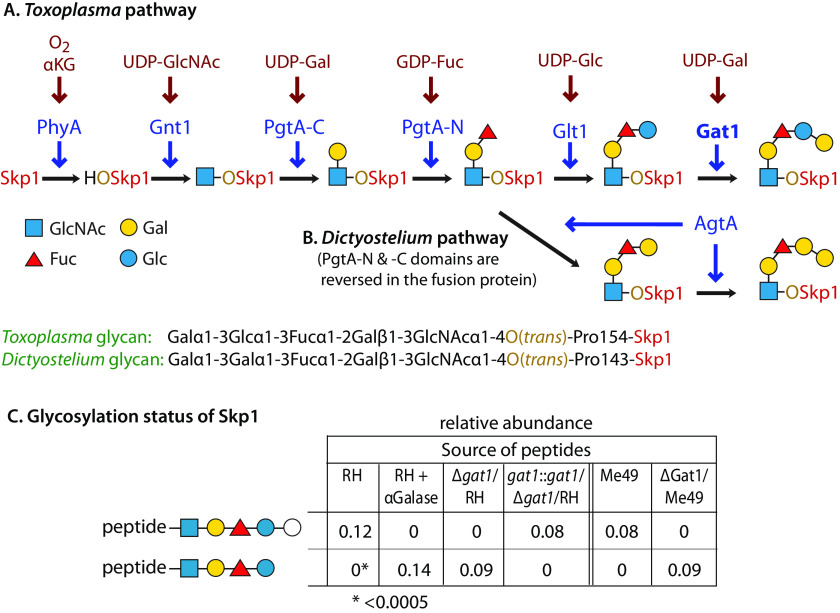
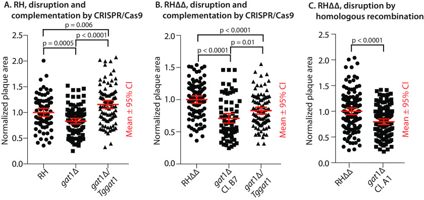
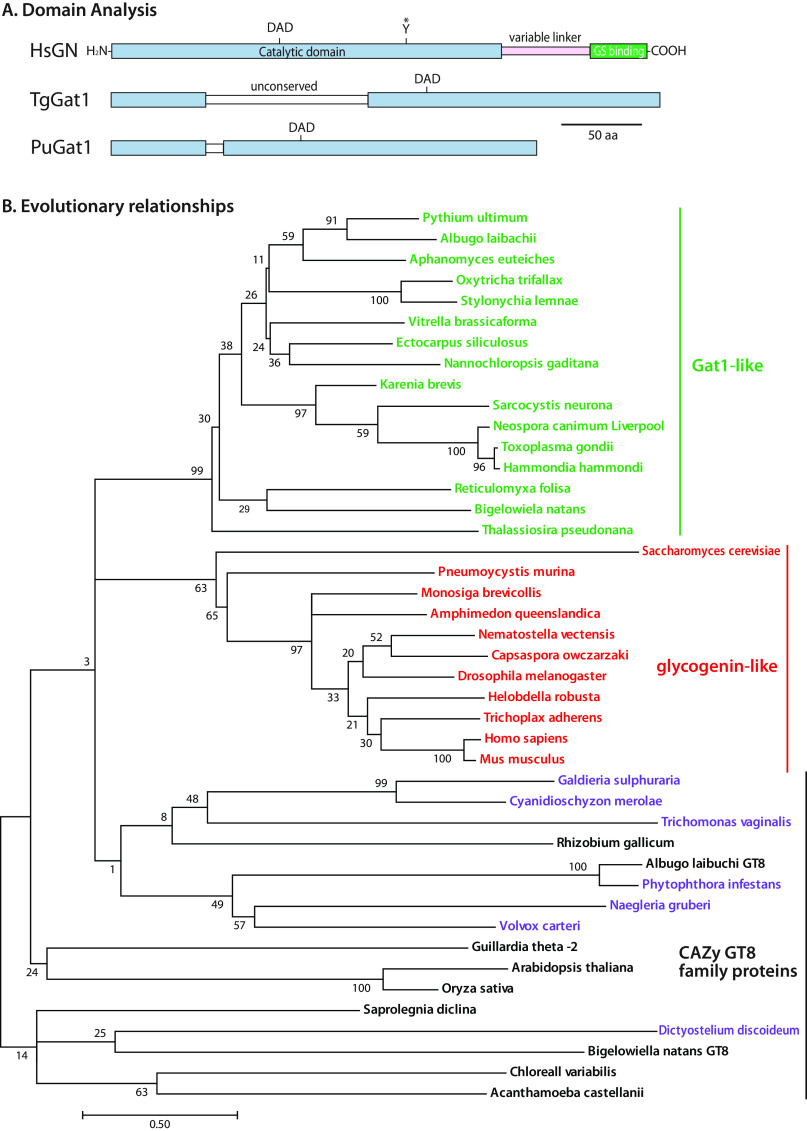
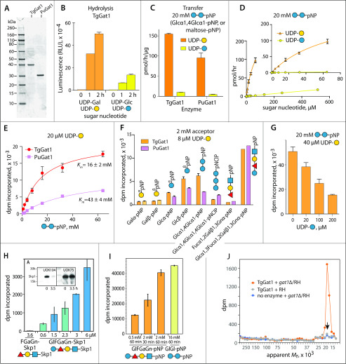
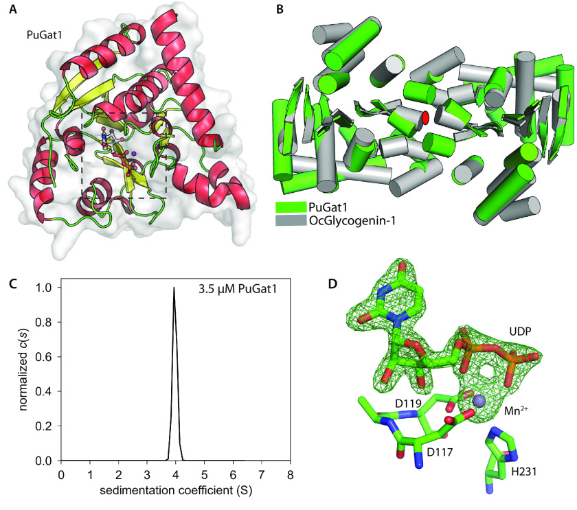
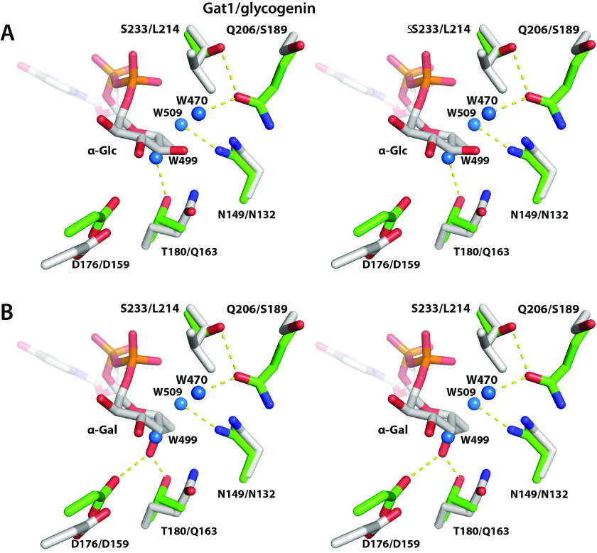
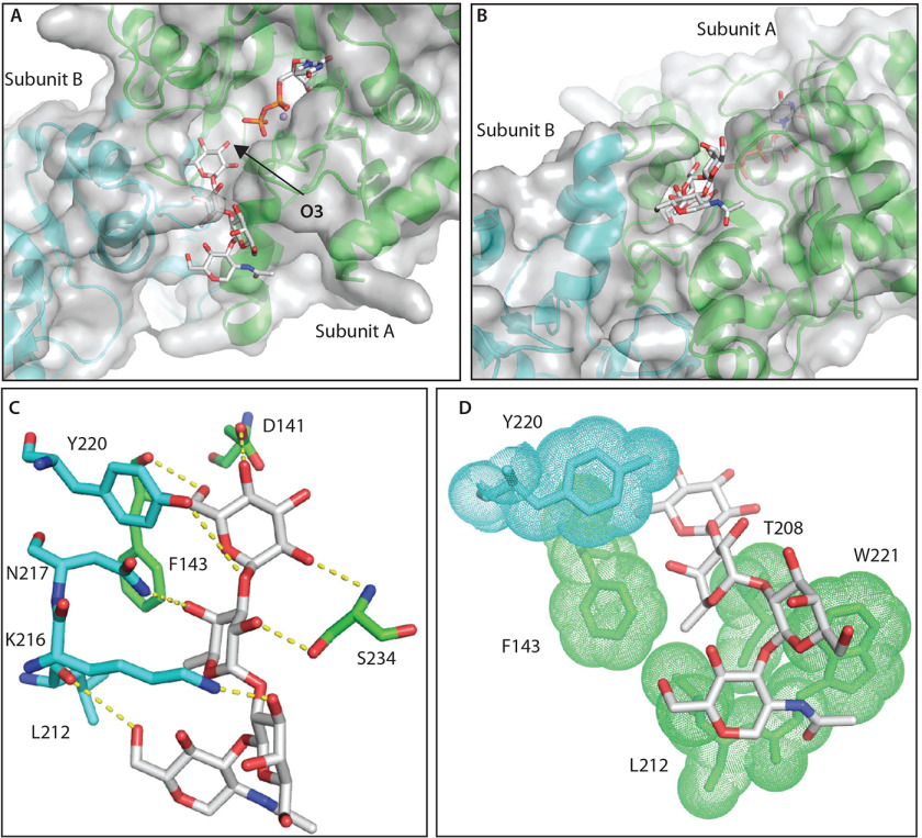
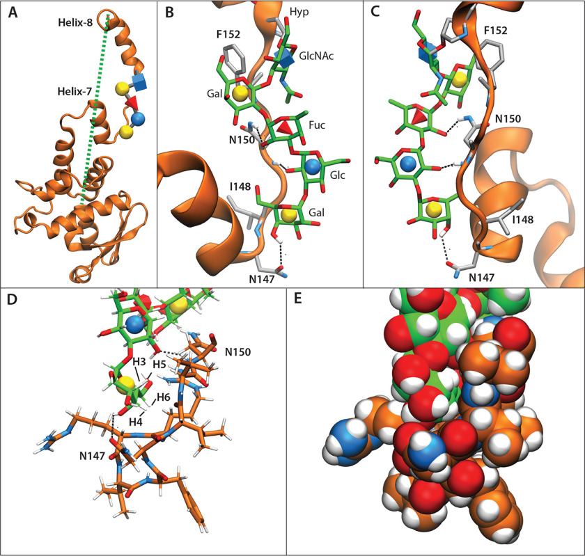
Skp1, a subunit of E3 Skp1/Cullin-1/F-box protein ubiquitin ligases, is modified by a prolyl hydroxylase that mediates O2 regulation of the social amoeba Dictyostelium and the parasite Toxoplasma gondii The full effect of hydroxylation requires modification of the hydroxyproline by a pentasaccharide that, in Dictyostelium, influences Skp1 structure to favor assembly of Skp1/F-box protein subcomplexes. In Toxoplasma, the presence of a contrasting penultimate sugar assembled by a different glycosyltransferase enables testing of the conformational control model. To define the final sugar and its linkage, here we identified the glycosyltransferase that completes the glycan and found that it is closely related to glycogenin, an enzyme that may prime glycogen synthesis in yeast and animals. However, the Toxoplasma enzyme catalyzes formation of a Galα1,3Glcα linkage rather than the Glcα1,4Glcα linkage formed by glycogenin. Kinetic and crystallographic experiments showed that the glycosyltransferase Gat1 is specific for Skp1 in Toxoplasma and also in another protist, the crop pathogen Pythium ultimum The fifth sugar is important for glycan function as indicated by the slow-growth phenotype of gat1Δ parasites. Computational analyses indicated that, despite the sequence difference, the Toxoplasma glycan still assumes an ordered conformation that controls Skp1 structure and revealed the importance of nonpolar packing interactions of the fifth sugar. The substitution of glycosyltransferases in Toxoplasma and Pythium by an unrelated bifunctional enzyme that assembles a distinct but structurally compatible glycan in Dictyostelium is a remarkable case of convergent evolution, which emphasizes the importance of the terminal α-galactose and establishes the phylogenetic breadth of Skp1 glycoregulation.

Keywords: E3 ubiquitin ligase; NMR; Pythium; SCF; Skp1; Toxoplasma; Toxoplasma gondii; X-ray crystallography; cytoplasmic glycosylation; glycogenin; glycosyltransferase; molecular dynamics; molecular dynamics simulation; nuclear magnetic resonance (NMR); post-translational modification; post-translational modification (PTM); s: X-ray crystallography.


Conflict of interest statement

Conflict of interest—The authors declare that they have no conflicts of interest with the contents of this article.

Figures

Figure 1.
Figure 1.
Gat1 is required for terminal α-galactosylation of Skp1 in parasites. A and B, schematic of Skp1 glycosylation pathway in Toxoplasma (14) (this work) and Dictyostelium (3), which modifies a single hydroxyproline associated with its F-box–binding region, depicted using CFG glycan symbols (19). The identity of Gat1 as the final enzyme in the Toxoplasma pathway and the nature of the final sugar are reported here. C, dependence of the terminal sugar on Gat1 and its characterization. Skp1 was immunoprecipitated from type 1 RH and type 2 ME49 strains and their genetic derivatives, and peptides generated by trypsinization were analyzed by nano-LC-MS. In addition, Skp1 from RH was treated with green coffee bean α-gal after trypsinization. The values represent the levels of pentasaccharide-peptide and tetrasaccharide-peptide levels detected, after normalization to all detected modification states of the peptide; note that the values have only partial relative meaning because of the low and varied detection efficiency of glycopeptides. As indicated, the detection threshold was <0.0005. The open circle for the terminal sugar of the pentasaccharide indicates that it was only known as a hexose at the time of the experiment. See Table S2 and Fig. S5 for primary data. Similar results were obtained in independent samples from RH and Δgat1/RH (not shown).
Figure 2.
Figure 2.
Parasite growth depends on Gat1. Parasites were plated at clonal density on two-dimensional monolayers of HFFs and allowed to invade, proliferate, lyse out, and reinfect neighboring fibroblasts. After 5.5 days, cultures were fixed, stained, and analyzed for the areas occupied by lysed fibroblasts (plaques). Data from each of three independent trials, which were each normalized to the parental strain, were merged for presentation. A, comparison of the type 1 RH strain before and after gat1 replacement using CRISPR/Cas9 and complementation with gat1 under control of its own promoter cassette in the uprt locus. B, comparison of RHΔΔ, gat1-2Δ/RHΔΔ, and the latter complemented with gat1 under control of a tubulin promoter in the uprt locus. C, comparison of gat1-1Δ/RHΔΔ, prepared by homologous recombination. Significance of differences in plaque areas between parasite strains was assessed by Student's t test. CI, confidence interval.
Figure 3.
Figure 3.
Gat1 is closely related to glycogenin sequences. A, domain analysis of Gat1 from T. gondii and P. ultimum compared with human glycogenin-1. B, the evolutionary history of the sequence of the Gat1 catalytic domain was inferred by using a maximum likelihood method. The tree with the highest log likelihood (−13,279.56) is shown. Gat1 and Gat1-like sequences are colored green, glycogenin and glycogenin-like sequences are in red, and characterized and other selected other CAZy GT8 sequences are in black, or in purple if predicted to reside in the secretory pathway rather than the cytoplasm. The percentage of trees in which the associated taxa clustered together is shown at each branch. Branch lengths are measured by the number of substitutions per site. See Figs. S6S8 for alignments.
Figure 4.
Figure 4.
Gat1 preferentially galactosylates the Skp1 glycan in vitro. A, recombinantly expressed and purified preparations of TgGat1 and PuGat1 were analyzed by SDS-PAGE and staining with Coomassie blue. B, temporal dependence of UDP-Gal and UDP-Glc hydrolysis. The averages and S.D. of three technical replicates are shown. A similar profile was observed with a different enzyme concentration. See Fig. S9E for a trial with higher enzyme concentrations. C, transferase activity utilizing 8 μm UDP-Gal or UDP-Glc toward 20 mm Glcα1,4Glcα-pNP (maltose-pNP) for TgGat1 and PuGat1. The averages and S.D. of two technical replicates are shown; similar profiles were in two independent assays with a different TgGat1 preparation. D, UDP-Gal and UDP-Glc concentration dependence of TgGat1 transferase activity toward 20 mm maltose-pNP. The averages and S.D. of two technical replicates are shown, and an independent trial with TgGat1 and PuGat1 against UDP-Gal is shown in Fig. S9F. E, maltose-pNP concentration dependence of TgGat1 and PuGat1 transferase activity from 20 μm UDP-Gal. The averages and S.D. of two technical replicates are shown. F, relative Gal-transferase activity of TgGat1 and PuGat1 toward different acceptors. The averages and S.D. of three technical replicates are shown. Similar results were obtained in three independent trials. G, effect of UDP-Glc concentration on the Gal-transferase activity of TgGat1. Reactions were incubated for 1 h. The averages and S.D. of two technical replicates are shown. H, Gal-transferase activity of TgGaT1 toward varied concentrations of GlFGaGn-Skp1, in the presence of 40 μm UDP-Gal (1 µCi) after a 1-h incubation. Data from independent preparations of TgSkp1 are colored in different shades. FGaGn-Skp1 is included for comparison. Error bars, S.D. of duplicate measurements. Inset, Western blots of the Skp1 preparations used, where FGaGn-Skp1, which is recognized specifically by pAb UOK104, is largely converted in a 3.5-h reaction using Glt1 and UDP-Glc to GlFGaGn-Skp1, which is recognized only by the pan-specific pAb UOK75. I, reactions with synthetic oligosaccharides conjugated to pNP were conducted in parallel using the same conditions. J, biochemical complementation to detect Gat1 substrates. Desalted S100 extracts of RH and gat1Δ/RH were reacted with recombinant Gat1 in the presence of UDP-[3H]Gal, and the product of the reaction was separated on an SDS-polyacrylamide gel, which was sliced into 40 bands for liquid scintillation counting. The migration position of Skp1 is marked with an arrow. See Fig. S9 (H and I) for trials using different strains.
Figure 5.
Figure 5.
Starch appears unaffected in gat1Δ parasites. To promote normal starch accumulation, rapidly proliferating tachyzoites (A) of the type II strain Me49 (RFP-expressing) and its gat1Δ derivative were induced to differentiate as slow-proliferating bradyzoite cysts (B) in human foreskin fibroblasts. Cultures were fixed and stained with periodic acid/Schiff's base to reveal starch as a purple adduct. Arrow in (A), parasitophorous vacuole containing dozens to hundreds of tachyzoites within a fibroblast. Arrow in (B), cyst containing dozens of slow-growing bradyzoites, as confirmed by labeling of the cyst wall with FITC-DBA lectin (not shown). Scale bars, 50 μm. Two independent trials yielded similar results.
Fig. 6.
Fig. 6.
Gat1 assembles a Galα1,3Glc linkage on the Skp1 tetrasaccharide. Shown is NMR analysis of the TgSkp1 pentasaccharide. [1-13C]Glcα1,3Fucα1,2Gal-β1,3GlcNAcα1-pNP was partially (30%) modified by TgGat1 or PuGat1 in the presence of UDP-[U-13C]Gal. A, one-dimensional 600-MHz 1H NMR spectrum. Magnification shows the region of 13C-anomeric carbons in the mixture of modified and unmodified tetrasaccharide. A cartoon diagram of the TgSkp1 pentasaccharide attached to pNP (Galα1,3Glcα1,3Fucα1,2Gal-β1,3GlcNAcα1-pNP) is shown at the top using CFG glycan symbols (19). B, 1H–13C HSQC, HMBC, and HSQC-TOCSY spectra demonstrating anomeric carbon to ring proton correlations. The Gal-H1/C1 doublet peaks were too weak to observe and are indicated by boxes. C, 1H–1H COSY and 1H–13C HMBC spectra. Identical results were obtained using [1-13C]GlFGaGn-pNP modified by PuGat1 (not shown). See Fig. S11 for a listing of chemical shift values.
Figure 7.
Figure 7.
Gat1 is structurally related to glycogenin. A, asymmetric unit of crystallized Gat1 from P. ultimum in complex with UDP and Mn2+. α-Helices are shown in red, β-strands in yellow, and loops in green, and the active site with bound ligand is boxed. B, PuGat1 forms a homodimer that closely superimposes on Oc-glycogenin-1 (PDB entry 1LL2). Cylinders, α-helices; arrows, β-sheets; red ellipse, 2-fold symmetry axis perpendicular to the page. C, sedimentation velocity data modeled as a continuous c(s) distribution (normalized to 1.0) yields an S value for 3.5 μm PuGat1 that is close to the predicted value for a stable dimer in solution. Fig. S12 shows that the dimer is stable down to at least 0.3 μm. D, UDP is coordinated in nearly identical fashion to that of glycogenin-1, based on the difference density map (FoFc) that was contoured at 5σ, calculated after omitting UDP and Mn2+ and subjecting the structure to simulated annealing. Octahedral coordination of Mn2+ is satisfied by the DXD motif, His-231, and UDP. The comparison with glycogenin-1 is illustrated in Fig. S13.
Figure 8.
Figure 8.
Active-site geometry explains Gat1's preference for UDP-Gal rather than UDP-Glc. Comparison of the sugar-binding pockets of PuGat1 and Oc-glycogenin-1 displayed as wall-eyed stereo view. A, the Glc moiety is modeled based on the Oc-glycogenin-1 crystal structure with its intact sugar nucleotide. B, the Gal moiety is modeled by flipping the stereochemistry of Glc at the C4′ position. PuGat1 and glycogenin-1 side chains are represented by green and gray sticks, respectively, the yellow dashes represent hydrogen bonds, and water molecules are represented by blue spheres.
Figure 9.
Figure 9.
Computational docking explains specificity of Gat1 for the Skp1-tetrasaccharide. A, top docking pose of the Skp1-tetrasaccharide in the PuGat1 active site and a groove formed by the dimer. B, 90° turn of the image shown in A. C, hydrogen-bonding interactions of the glycan. D, hydrophobic packing of the sugar faces and fucose-methyl (in sticks) with nonpolar surfaces (sticks/dots) of Gat1 subunit A (green) and B (cyan).
Figure 10.
Figure 10.
Packing of the glycan with Skp1 can explain F-box–binding site conformation. T. gondii GaGlFGaGn-Skp1 was subjected to six 250-ns all-atom molecular dynamics simulations. A, frame representative of the glycan-protein interaction and associated helix-8 extension, from a simulation (Equil-1; see Fig. S14E) in which the glycan was pre-equilibrated for 50 ns prior to the start of the simulation. The dotted green line refers to the distance from the C terminus to the center of mass of residues 1-136 and ranged from 18 to 61 Å. B, zoom-in of A depicting the glycan (carbon atoms in green) and amino acids (carbon atoms in gray) described in Table 2. Dotted black lines depict hydrogen bonds contributing to the polar energies described in Table 2. C, the back side of B. D and E, packing of terminal sugars against the polypeptide. Carbon atoms of the peptide are in orange. D, glycan and peptide represented as sticks. E, as in D, with glycan and peptide represented by spheres.

References

    1. Islam M. S., Leissing T. M., Chowdhury R., Hopkinson R. J., and Schofield C. J. (2018) 2-Oxoglutarate-dependent oxygenases. Annu. Rev. Biochem. 87, 585–620 10.1146/annurev-biochem-061516-044724 - DOI - PubMed
    1. Pugh C. W., and Ratcliffe P. J. (2017) New horizons in hypoxia signaling pathways. Exp. Cell Res. 356, 116–121 10.1016/j.yexcr.2017.03.008 - DOI - PMC - PubMed
    1. West C. M., and Blader I. J. (2015) Oxygen sensing by protozoans: how they catch their breath. Curr. Opin. Microbiol. 26, 41–47 10.1016/j.mib.2015.04.006 - DOI - PMC - PubMed
    1. Willems A. R., Schwab M., and Tyers M. (2004) A hitchhiker's guide to the cullin ubiquitin ligases: SCF and its kin. Biochim. Biophys. Acta 1695, 133–170 10.1016/j.bbamcr.2004.09.027 - DOI - PubMed
    1. West C. M., van der Wel H., and Wang Z. A. (2007) Prolyl 4-hydroxylase-1 mediates O2 signaling during development of Dictyostelium. Development 134, 3349–3358 10.1242/dev.000893 - DOI - PubMed

Publication types

MeSH terms

LinkOut - more resources