. 2021 Apr 1;106(4):1034-1046.
doi: 10.3324/haematol.2019.244020.

Venetoclax combines synergistically with FLT3 inhibition to effectively target leukemic cells in FLT3-ITD+ acute myeloid leukemia models

Affiliations

Venetoclax combines synergistically with FLT3 inhibition to effectively target leukemic cells in FLT3-ITD+ acute myeloid leukemia models

Raghuveer Singh Mali et al. Haematologica. .

Abstract

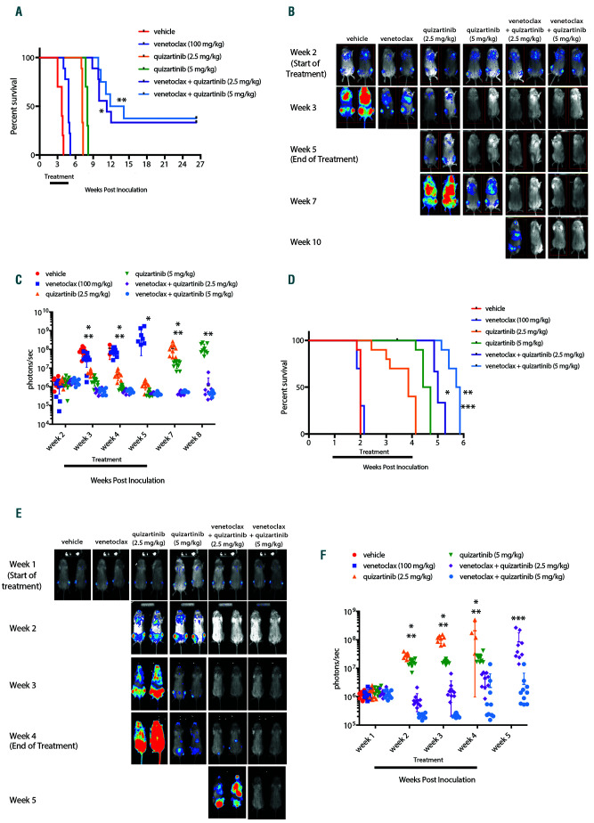
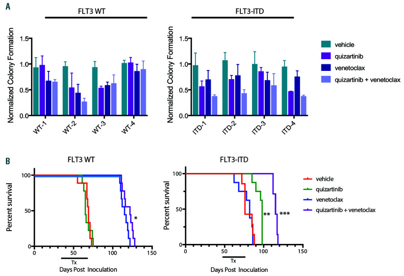
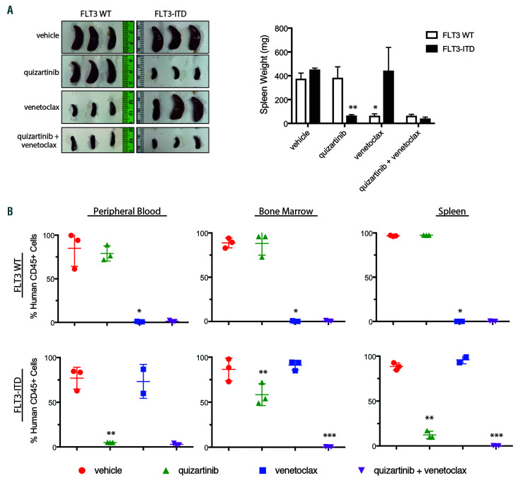
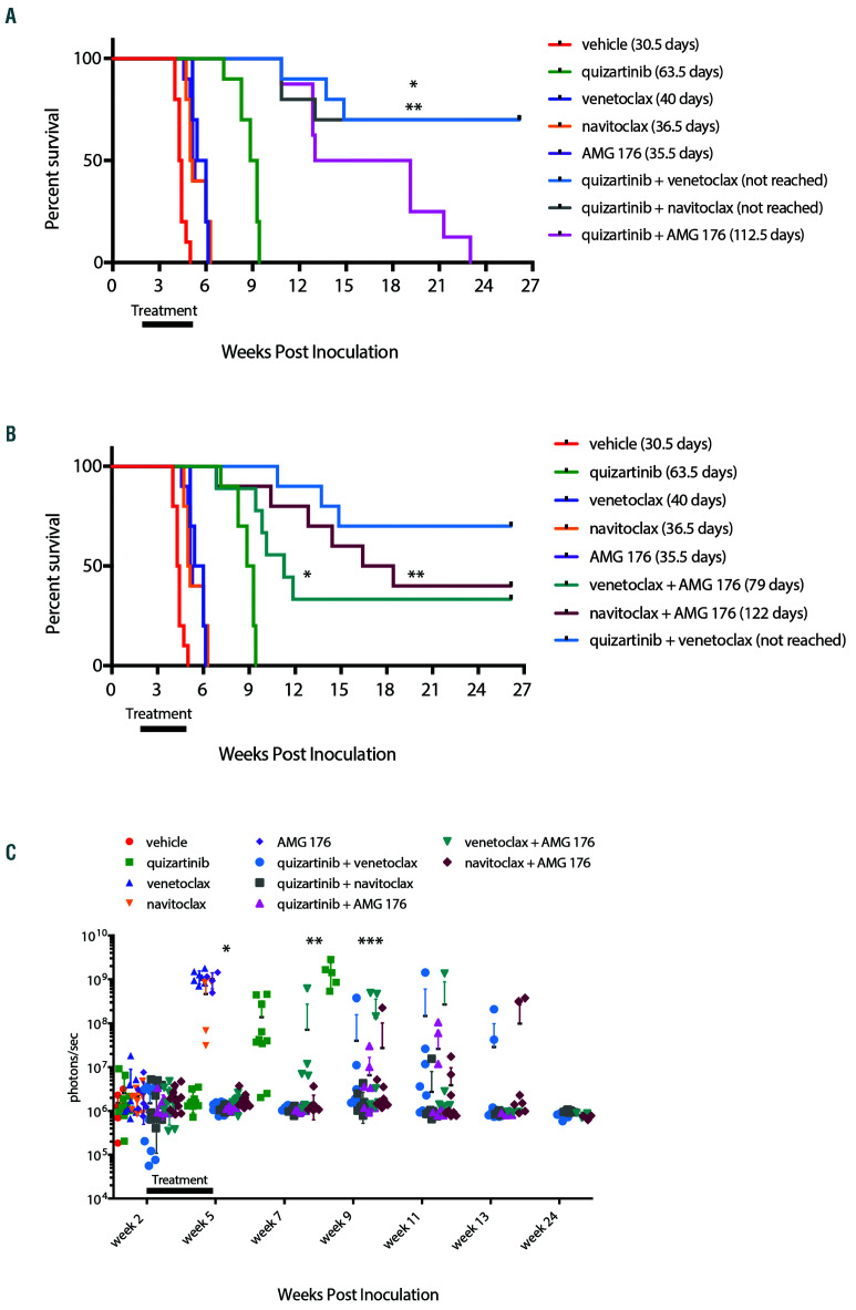
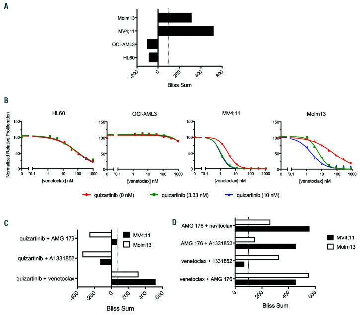
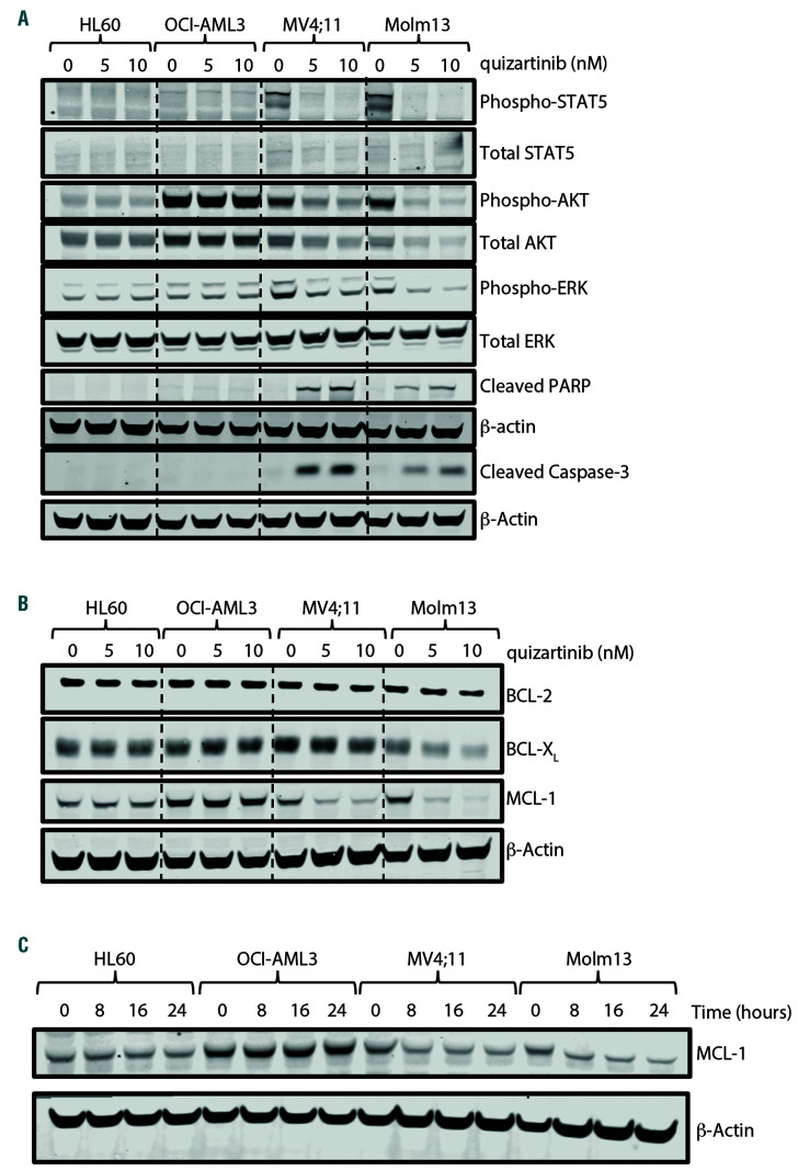
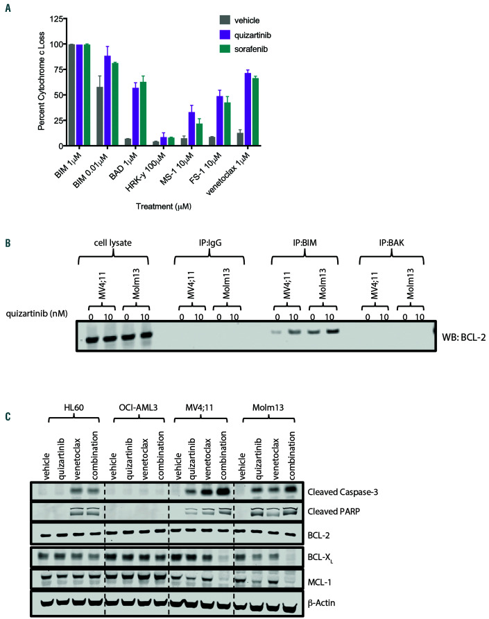
FLT3 internal tandem duplication (FLT3-ITD) mutations account for ~25% of adult acute myeloid leukemia cases and are associated with poor prognosis. Venetoclax, a selective BCL-2 inhibitor, has limited monotherapy activity in relapsed/refractory acute myeloid leukemia with no responses observed in a small subset of FLT3-ITD+ patients. Further, FLT3-ITD mutations emerged at relapse following venetoclax monotherapy and combination therapy suggesting a potential mechanism of resistance. Therefore, we investigated the convergence of FLT3-ITD signaling on the BCL-2 family proteins and determined combination activity of venetoclax and FLT3-ITD inhibition in preclinical models. In vivo, venetoclax combined with quizartinib, a potent FLT3 inhibitor, showed greater anti-tumor efficacy and prolonged survival compared to monotherapies. In a patient-derived FLT3-ITD+ xenograft model, cotreatment with venetoclax and quizartinib at clinically relevant doses had greater anti-tumor activity in the tumor microenvironment compared to quizartinib or venetoclax alone. Use of selective BCL-2 family inhibitors further identified a role for BCL-2, BCL-XL and MCL-1 in mediating survival in FLT3-ITD+ cells in vivo and highlighted the need to target all three proteins for greatest anti-tumor activity. Assessment of these combinations in vitro revealed synergistic combination activity for quizartinib and venetoclax but not for quizartinib combined with BCL-XL or MCL-1 inhibition. FLT3-ITD inhibition was shown to indirectly target both BCL-XL and MCL-1 through modulation of protein expression, thereby priming cells toward BCL-2 dependence for survival. These data demonstrate that FLT3-ITD inhibition combined with venetoclax has impressive anti-tumor activity in FLT3-ITD+ acute myeloid leukemia preclinical models and provides strong mechanistic rational for clinical studies.


Figures

Figure 1.
Figure 1.
Venetoclax combined with quizartinib prolongs survival and reduces tumor burden in FLT3-ITD+ xenograft models. NOD/SCID/IL-2Rγnull (NSG) mice were engrafted with luciferase expressing MV4;11 or Molm13 cells and leukemic cell engraftment was confirmed by bioluminescence imaging (BLI) and treatment began 14 days post-inoculation for the MV4;11 model and 7 days post-inoculation in the Molm13 model. Mice were treated orally with 100 mg/kg venetoclax, 2.5 mg/kg or 5 mg/kg quizartinib, or the combination once daily for 21 continuous days and overall survival and disease burden was assessed. (A) Kaplan- Meier survival curve for MV4;11-engrafted mice. N=8-10 animals/group and median survival and statistical significance were determined by log-rank test: *P<0.0001 for venetoclax + 2.5 mg/kg quizartinib vs. 2.5 mg/kg quizartinib; **P<0.0001 for venetoclax + 5 mg/kg quizartinib vs. 5 mg/kg quizartinib. (B) Representative BLI for MV4;11-engrafted mice for each group at indicated time point. (C) Quantitation of the BLI signal from MV4;11-engrafted mice in each group at indicated time post-inoculation: *P<0.0001, P=0.0002, P=0.001 and P=0.0004 for venetoclax + 2.5 mg/kg quizartinib vs. 2.5 mg/kg quizartinib for week 3, 4, 5 and 7, respectively, by unpaired t-test for each time point; **P=0.0004, P=0.0247, P<0.0001 and P=0.0001 for venetoclax + 5 mg/kg quizartinib vs. 5 mg/kg quizartinib for week 3, 4, 7 and 8, respectively, by unpaired t-test for each time point. (D) Kaplan-Meier curve for Molm13-engrafted mice. N=10 animals/group and survival and statistical significance were determined by log-rank test: *P<0.0001 for venetoclax + 2.5 mg/kg quizartinib vs. 2.5 mg/kg quizartinib; **P<0.0001 for venetoclax + 5 mg/kg quizartinib vs. 5 mg/kg quizartinib; and ***P<0.0001 for venetoclax + 5 mg/kg quizartinib vs. venetoclax + 2.5 mg/kg quizartinib. (E) Representative BLI for Molm13-engrafted mice at indicated time point. (F) Quantitation of BLI signal from Molm13-engrafted mice at indicated time point post-inoculation: *P<0.0001, P<0.0001 and P=0.0086 for venetoclax + 2.5 mg/kg quizartinib vs. 2.5 mg/kg quizartinib for week 2, 3 and 4, respectively, by unpaired t test for each timepoint; **P<0.0001 for venetoclax + 5 mg/kg quizartinib vs. 5 mg/kg quizartinib for week 2, 3 and 4 by unpaired t-test for each timepoint; and ***P=0.0129 for venetoclax + 5 mg/kg quizartinib vs. venetoclax + 2.5 mg/kg quizartinib at week 5 by unpaired t-test.
Figure 2.
Figure 2.
Venetoclax combined with quizartinib has greater anti-tumor efficacy against primary FLT3-ITD+ acute myeloid leukemia models. (A) Primary patient samples were treated with 20 nM quizartinib, 50 nM venetoclax or the combination in methylcellulose in triplicate for 14 days and colony forming units were counted. Data were normalized to the median colony number for the vehicle-treated cells for each sample and plotted as mean + standard deviation. (B) NOD/SCID/IL-2Rγnull (NSG) mice were engrafted with primary samples from Fms-like tyrosine kinase 3 wild-type (FLT3-WT) or FLT3 internal tandem duplication (FLT3-ITD+) patients and treated orally with 5 mg/kg quizartinib, 100 mg/kg venetoclax or the combination once daily for 28 days and efficacy was assessed. Kaplan-Meier survival curve for FLT3-WT and FLT3-ITD+ models. N=7-9 animals/group and median survival and statistical significance were determined by log-rank test: *P<0.0001 for venetoclax vs. vehicle or quizartinib in FLT3-WT model. **P=0.0004 for quizartinib vs. vehicle or venetoclax and ***P=0.0003 for quizartinib + venetoclax vs. quizartinib in the FLT3-ITD+ model.
Figure 3.
Figure 3.
Venetoclax combined with quizartinib to reduce disease burden in the tumor microenvironment of patient-derived FLT3-ITD+ xenograft models. NOD/SCID/IL-2Rγnull (NSG) mice were engrafted with primary samples from Fms-like tyrosine kinase 3 wild-type (FLT3-WT) or FLT3 internal tandem duplication (FLT3- ITD+) patients and were treated orally with 5 mg/kg quizartinib, 100 mg/kg venetoclax or the combination once daily for 28 days. (A) Spleen images and spleen weight at the end of 28 days of dosing. Data is represented as average + standard deviation (n=3). *P=0.0004 for venetoclax vs. quizartinib for FLT3-WT model and **P=0.0068 for quizartinib vs. venetoclax for FLT3-ITD+ model by one-way ANOVA with Tukey post hoc test. (B) Percentage of human CD45+ cells in peripheral blood, bone marrow and spleen at the end of dosing. Data is represented as average + standard deviation (n=3). *P<0.0001 for venetoclax vs. quizartinib in the FLT3-WT model in the peripheral blood, bone marrow and spleen; **P=0.0005, P=0.01 and P<0.0001 for quizartinib vs. venetoclax for peripheral blood, bone marrow and spleen, respectively, for FLT3-ITD+ model; and *** P=0.0002 and P=0.0159 for quizartinib + venetoclax vs. quizartinib for bone marrow and spleen, respectively, for FLT3-ITD+ model by one-way ANOVA with Tukey post hoc test.
Figure 4.
Figure 4.
BCL-2, BCL-XL and MCL-1 contribute to survival of FLT3-ITD+ cells in vivo. NOD/SCID/IL-2Rγnull (NSG) mice were engrafted with luciferase expressing MV4;11 cells and leukemic engraftment was confirmed by BLI and treatment began 14 days post inoculation. Mice were treated orally with 5 mg/kg quizartinib, 100 mg/kg venetoclax, 100 mg/kg navitoclax or 10 mg/kg AMG 176 as single agents or in combinations as indicated once daily for 21 days. All treatment groups were run concurrently, however for clarity survival data has been split between panels A and B and vehicle, quizartinib, venetoclax, navitoclax and AMG 176 single agent groups have been plotted in both panels for reference. (A) Kaplan-Meier survival curves for quizartinib in combination with AMG 176, venetoclax or navitoclax. N=8-10 animals/group. Median survival and statistics were determined by log-rank test: *P=0.0059 for quizartinib + venetoclax vs. quizartinib + AMG 176; and **P=0.0095 for quizartinib + navitoclax vs. quizartinib + AMG 176. (B) Kaplan-Meier survival curves for AMG 176 in combination with venetoclax or navitoclax. N=8-10 animals/ group. Median survival and statistics were determined by log-rank test: *P=0.0008 for venetoclax + AMG 176 vs. quizartinib; and **P=0.0002 for navitoclax + AMG 176 vs. quizartinib. (C) Quantitation of the bioluminescence imaging (BLI) signal from MV4;11-engrafted mice in each group at indicated time post-inoculation: *P<0.005 for quizartinib single agent and all combinations vs. venetoclax, AMG 176 or navitoclax single agents at week 5 by one-way ANOVA with Tukey post-test; **P<0.05 for quizartinib + venetoclax, quizartinib + navitoclax, quizartinib + AMG 176 and navitoclax + AMG 176 vs. quizartinib single agent on week 7 by one-way ANOVA with Dunnett post-test; ***P=0.0001 for quizartinib + venetoclax, quizartinib + navitoclax, quizartinib + AMG 176, venetoclax + AMG 176 and navitoclax + AMG 176 vs. single agent quizartinib at week 9 by one-way ANOVA with Dunnett post-test.
Figure 5.
Figure 5.
Venetoclax synergistically combined with quizartinib in FLT3-ITD+ cell lines. (A) Cell lines were treated for 48 hours with venetoclax, quizartinib or the combination at indicated concentrations. ATP content was determined by CellTiter-Glo and Bliss sums were calculated and plotted for each cell line. Bliss sum of >100 is highly synergistic. (B) Cell lines were treated for 48 hours with quizartinib, venetoclax or the combination as indicated and cell viability was assessed by CellTiter- Glo. Values are normalized to the average of the untreated samples for each cell line. (C) Fms-like tyrosine kinase 3 (FLT3) internal tandem duplication (FLT3-ITD+) cell lines were treated for 48 hours with combinations of quizartinib and venetoclax, AMG 176 or A1331852 as indicated. ATP content was determined by CellTiter- Glo and Bliss sums were calculated and plotted for each cell line. (D) FLT3-ITD+ cell lines were treated for 48 hours with combinations of venetoclax, AMG 176, A1331852 or navitoclax as indicated. ATP content was determined by CellTiter-Glo and Bliss sums were calculated and plotted for each cell line.
Figure 6.
Figure 6.
FLT3-ITD signaling regulates the expression of BCL-XL and MCL-1 in vitro. Fms-like tyrosine kinase 3 wild-type (FLT3-WT) (HL60 and OCI-AML3) and FLT3 internal tandem duplication (FLT3-ITD+) (MV4;11 and Molm13) cell lines treated for 24 hours with indicated concentration of quizartinib and cell lysates analyzed by western blot for (A) FLT3-ITD downstream effector proteins and (B) BCL-2 family proteins as indicated. (C) Cell lines were treated with 10 nM quizartinib for indicated time and MCL-1 expression was assessed in whole cell lysate by western blot.
Figure 7.
Figure 7.
FLT3-ITD inhibition primed cells to BCL-2 dependence. (A) MV4;11 cells were pre-treated for 6 hours with vehicle, 5 nM quizartinib or 20 nM sorafenib and depletion of intracellular cytochrome c was determined following 1 hour exposure to BIM, BAD, HRK, MS- 1 and FS-1 peptides or venetoclax at the indicated concentrations by flow cytometry. Data represents average ± standard deviation within the experiment. (B) MV4;11 and Molm13 cells were treated with 10 nM quizartinib for 24 hours followed by immunoprecipitation for BIM or BAK as indicated followed by Western blot analysis for BCL-2. (C) Cell lines were treated with 100 nM venetoclax, 10 nM quizartinib or the combination for 24 hours and cell lysates were assessed by Western blot for BCL- 2 family proteins, cleaved caspase-3 and cleaved PARP as indicated. Data represents two independent experiments. FLT3-ITD: Fms-like tyrosine kinase 3 internal tandem duplication.

References

    1. Levis M, Small D. FLT3: ITDoes matter in leukemia. Leukemia. 2003;17(9):1738-1752. - PubMed
    1. Yamamoto Y, Kiyoi H, Nakano Y, et al. . Activation mutation of D835 within the activation loop of FLT3 in human hematologic malignancies. Blood. 2001;97(8):2434-2439. - PubMed
    1. Choudhary C, Muller-Tidow C, Berdel WE, Serve H. Signal transduction of oncogenic Flt3. Int J Hematol. 2005;82(2):93-99. - PubMed
    1. Hayakawa F, Towatari M, Kiyoi H, et al. . Tandem-duplicated Flt3 constitutively activates STAT5 and MAP kinase and introduces autonomous cell growth in IL3- dependent cell lines. Oncogene. 2000; 19:624-631. - PubMed
    1. Mizuki M, Fenski R, Halfter H, et al. . Flt3 mutations from patients with acute myeloid leukemia induce transformation of 32D cells mediated by the Ras and STAT5 pathways. Blood. 2000;96(12):3907-3914. - PubMed

MeSH terms