. 2021 Jun 1;106(6):1714-1724.
doi: 10.3324/haematol.2019.245860.

Molecular profiling reveals a hypoxia signature in breast implant-associated anaplastic large cell lymphoma

Affiliations

Molecular profiling reveals a hypoxia signature in breast implant-associated anaplastic large cell lymphoma

Naoki Oishi et al. Haematologica. .

Abstract

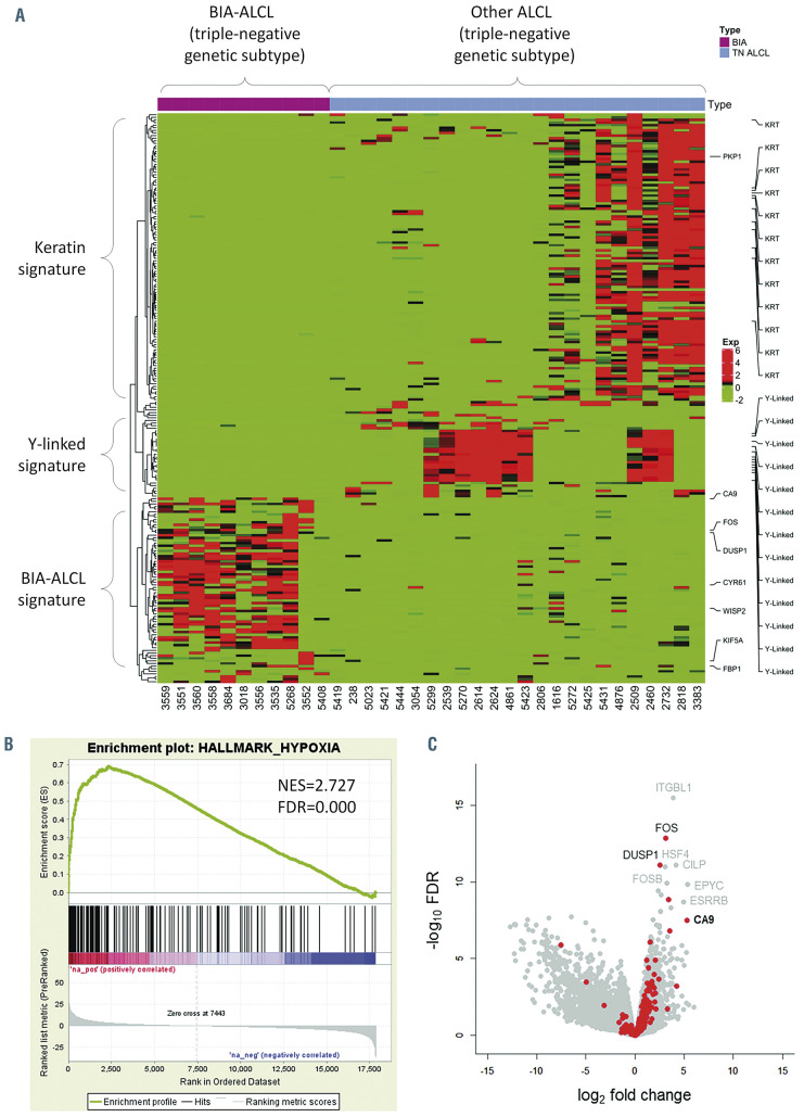
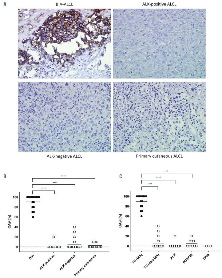
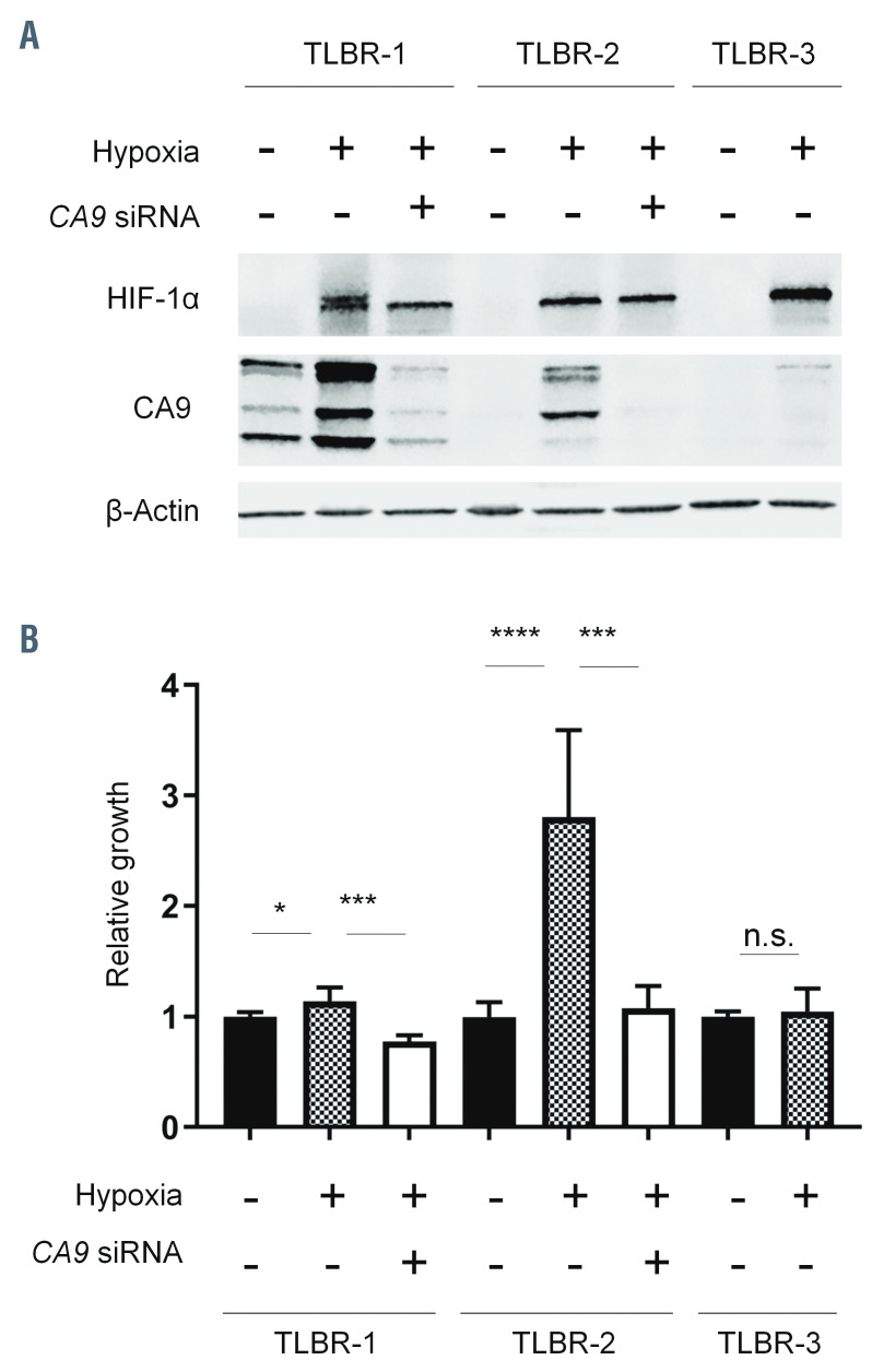
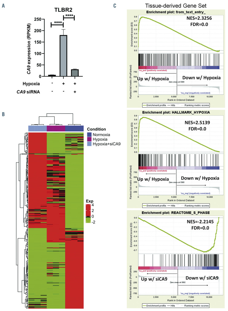
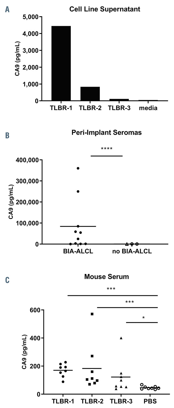
Breast implant-associated anaplastic large cell lymphoma (BIA-ALCL) is a recently characterized T-cell malignancy that has raised significant patient safety concerns and led to worldwide impact on the implants used and clinical management of patients undergoing reconstructive or cosmetic breast surgery. Molecular signatures distinguishing BIA-ALCL from other ALCLs have not been fully elucidated and classification of BIA-ALCL as a WHO entity remains provisional. We performed RNA sequencing and gene set enrichment analysis comparing BIA-ALCLs to non-BIA-ALCLs and identified dramatic upregulation of hypoxia signaling genes including the hypoxia-associated biomarker CA9 (carbonic anyhydrase-9). Immunohistochemistry validated CA9 expression in all BIA-ALCLs, with only minimal expression in non-BIA-ALCLs. Growth induction in BIA-ALCL-derived cell lines cultured under hypoxic conditions was proportional to up-regulation of CA9 expression, and RNA sequencing demonstrated induction of the same gene signature observed in BIA-ALCL tissue samples compared to non-BIA-ALCLs. CA9 silencing blocked hypoxia-induced BIA-ALCL cell growth and cell cycle-associated gene expression, whereas CA9 overexpression in BIA-ALCL cells promoted growth in a xenograft mouse model. Furthermore, CA9 was secreted into BIA-ALCL cell line supernatants and was markedly elevated in human BIA-ALCL seroma samples. Finally, serum CA9 concentrations in mice bearing BIA-ALCL xenografts were significantly elevated compared to control serum. Together, these findings characterize BIA-ALCL as a hypoxia-associated neoplasm, likely attributable to the unique microenvironment in which it arises. These data support classification of BIA-ALCL as a distinct entity and uncover opportunities for investigating hypoxia-related proteins such as CA9 as novel biomarkers and therapeutic targets in this disease.


Figures

Figure 1.
Figure 1.
Breast implant-associated anaplastic large cell lymphomas show upregulation of hypoxia-associated genes. (A) Heatmap of genes differentially expressed between breast implant-associated (BIA) anaplastic large cell lymphomas (ALCL) and other ALCL of triple-negative (TN) genetic subtype (lacking rearrangements of ALK, DUSP22 and TP63). A distinct signature of genes overexpressed in BIA-ALCL is seen. Signatures in subgroups of non-BIA ALCL include a keratin (KRT) signature seen in biopsies at epithelial sites (skin and tongue) and a Y-linked signature seen in male patients. See also Online Supplementary Figure S1. (B) Gene set enrichment analysis shows upregulation of genes associated with hypoxia in BIA-ALCL. See also Online Supplementary Table S2. NES: normalized enrichment score; FDR: false discovery rate. (C) A volcano plot indicating differentially expressed genes between BIA-ALCL and other TN ALCL. Hypoxia-related genes from HALLMARK HYPOXIA and SEMENZA HIF1 TARGETS gene sets are indicated in red.
Figure 2.
Figure 2.
Breast implant-associated anaplastic large cell lymphomas consistently express CA9. (A) Representative microscopic images of immunohistochemistry for CA9 in breast implant-associated (BIA) anaplastic large cell lymphoma (ALCL), systemic ALK-positive ALCL, systemic ALK-negative ALCL, and primary cutaneous ALCL (40× original magnification). (B) BIA-ALCL show significantly higher CA9 expression than other forms of ALCL. (C) The increased expression of CA9 in BIA-ALCL is independent of genetic subtype. All BIA-ALCL tested have triple-negative (TN) genetics (lacking rearrangements of ALK, DUSP22, and TP63). BIA-ALCL show significantly higher CA9 expression than ALCL with any of these rearrangements, as well as TN non-BIA-ALCL. ***P<0.001; ****P<0.0001.
Figure 3.
Figure 3.
CA9 expression and growth of breast implant-associated anaplastic large cell lymphoma cell lines. (A) TLBR-1, -2, and -3 breast implant-associated (BIA) anaplastic large cell lymphoma (ALCL) cell lines show distinct patterns of CA9 expression under normoxic and hypoxic conditions. HIF-1α serves as a positive control for hypoxia. TLBR-1 shows constitutive CA9 expression under normoxic conditions, which is further enhanced by hypoxia. TLBR-2 lacks constitutive CA9 expression but CA9 is induced by hypoxia (canonical hypoxia response). TLBR-3 shows minimal hypoxia-induced CA9 expression. The effects of siRNAmediated CA9 silencing are shown. Representative data from three independent experiments. (B) Growth of TLBR-1, -2, and -3 cells mirrors CA9 expression. TLBR-1 cells, which constitutively express CA9, show only slight growth induction by hypoxia. Growth is inhibited by CA9 silencing. TLBR-2 cells, which show a canonical hypoxia response, have marked hypoxia-induced growth which is almost completely reversed by CA9 silencing. Hypoxia does not induce either growth or CA9 expression in TLBR-3 cells. *P<0.05; ***P<0.001; ****P<0.0001; n.s., not statistically significant.
Figure 4.
Figure 4.
Gene signatures associated with hypoxia and CA9 expression in breast implant-associated anaplastic large cell lymphoma cells. (A) CA9 mRNA expression is induced by hypoxia and inhibited by CA9 siRNA in TLBR-2 breast implant-associated (BIA) anaplastic large cell lymphoma (ALCL) cells. RPKM: reads per kilobase per million mapped reads; ****P<0.0001. (B) Heatmap showing relative gene expression in TLBR-2 for each of the three conditions shown in panel A. RNA sequencing was performed in triplicate for each condition. (C) Top panel: TLBR-2 cells cultured under hypoxic conditions show significant enrichment for the set of genes overexpressed in BIA- versus non-BIA-ALCL tissue samples (“tissue-derived gene set,” defined as log2FC >1 and FDR ≤0.05; cf. Figure 1A). Middle panel: the HALLMARK HYPOXIA gene set identified by gene set enrichment analysis in BIA- versus non-BIA-ALCL tissue samples is also significantly enriched in hypoxic TLBR- 2 cells. See also Online Supplementary Table S2. Bottom panel: hypoxic TLBR-2 cells treated with CA9 siRNA show marked depletion of REACTOME S PHASE and other gene sets related to cell cycle and MYC targets. See also Online Supplementary Table S3. FC: fold change; NES: normalized enrichment score; FDR: false discovery rate.
Figure 5.
Figure 5.
CA9 accelerates breast implant-associated anaplastic large cell lymphoma growth in a mouse xenograft model. (A) Mice inoculated with TLBR-3 breast implant-associated anaplastic large cell lymphoma cells stably transduced with a CA9 lentiviral vector develop palpable tumors faster than mice inoculated with cells transduced with vector control. (B) Mice inoculated with TLBR-3 cells overexpressing CA9 develop larger tumors than mice inoculated with control-transduced TLBR-3 cells. (C) Overall survival is shorter in mice bearing CA9-overexpressing TLBR-3 tumors than in those bearing control-transduced tumors.
Figure 6.
Figure 6.
CA9 as a candidate biomarker in breast implant-associated anaplastic large cell lymphoma. (A) TLBR-1, -2, and -3 cell lines secrete CA9 into culture supernatant proportionally to their cellular expression, as determined by western blot (cf. Figure 3A). Data represent three replicates measured by CA9 enzyme-linked immunosorbent assay. (C) Peri-implant seroma samples involved by breast implant-associated (BIA) anaplastic large cell lymphoma (ALCL) have significantly higher CA9 concentrations than those not involved by BIA-ALCL. (C) Serum samples obtained from mice bearing 1000 mm subcutaneous TLBR-1, - 2, and -3 tumors have significantly higher CA9 concentrations than those from non-tumor-bearing mice. *P<0.05; **P<0.01; ***P<0.001; ****P<0.0001 (Mann-Whitney test). PBS: phosphate-buffered saline.

Comment in

References

    1. Xing X, Feldman AL. Anaplastic large cell lymphomas: ALK positive, ALK negative, and primary cutaneous. Adv Anat Pathol. 2015;22(1):29-49. - PubMed
    1. Feldman AL, Harris NL, Stein H, et al. . Breast implant-associated anaplastic large cell lymphoma. Swerdlow SH, Campo E, Harris NL, et al.., eds. WHO Classification of Tumours of Haematopoietic and Lymphoid Tissues. Revised 4th ed. Lyon: International Agency for Research on Cancer, 2017:421-422.
    1. Miranda RN, Aladily TN, Prince HM, et al. . Breast implant-associated anaplastic largecell lymphoma: long-term follow-up of 60 patients. J Clin Oncol. 2014;32(2):114-120. - PMC - PubMed
    1. Ferrufino-Schmidt MC, Medeiros LJ, Liu H, et al. . Clinicopathologic features and prognostic impact of lymph node involvement in patients with breast implant-associated anaplastic large cell lymphoma. Am J Surg Pathol. 2018;42(3):293-305. - PMC - PubMed
    1. Clemens MW, Medeiros LJ, Butler CE, et al. . Complete surgical excision is essential for the management of patients with breast implant-associated anaplastic large-cell lymphoma. J Clin Oncol. 2016;34(2):160-168. - PMC - PubMed

Publication types