Randomized Controlled Trial
. 2020 May;41(5):758-765.
doi: 10.3174/ajnr.A6560.

Methylphenidate Effects on Cortical Thickness in Children and Adults with Attention-Deficit/Hyperactivity Disorder: A Randomized Clinical Trial

Affiliations
Randomized Controlled Trial

Methylphenidate Effects on Cortical Thickness in Children and Adults with Attention-Deficit/Hyperactivity Disorder: A Randomized Clinical Trial

K B Walhovd et al. AJNR Am J Neuroradiol. 2020 May.

Abstract

Background and purpose: Although methylphenidate is frequently used to treat children with attention-deficit/hyperactivity disorder, it is currently unknown how methylphenidate affects brain development. In a randomized controlled trial, we investigated whether the cortical effects of methylphenidate are modulated by age.

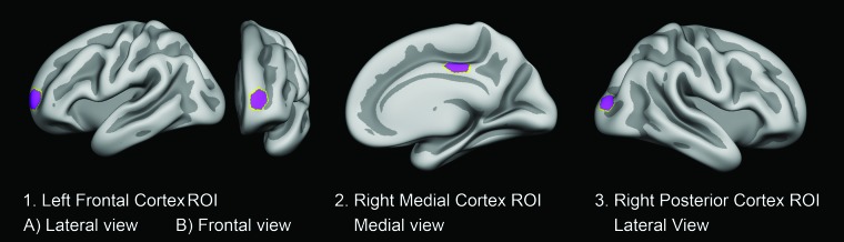
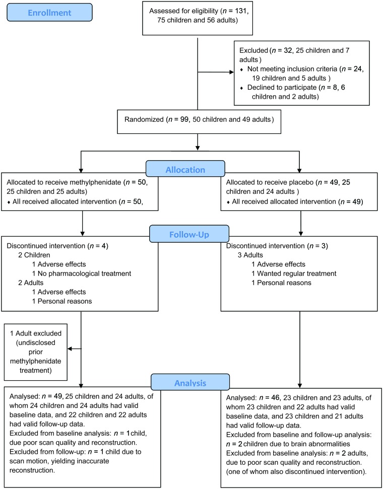
Materials and methods: Between June 1, 2011, and June 15, 2015, we conducted a randomized, double-blind, placebo-controlled trial (Effects of Psychotropic Drugs on Developing Brain-Methylphenidate) in 99 males with attention-deficit/hyperactivity disorder (according to Diagnostic and Statistical Manual of Mental Disorders, 4th Edition, criteria) from referral centers in the greater Amsterdam area in the Netherlands. The trial was registered on March 24, 2011 (identifier NL34509.000.10) and subsequently at the Netherlands National Trial Register (identifier NTR3103). Participants (first enrolled October 13, 2011) were 10-12 years or 23-40 years of age and randomized to treatment with either methylphenidate or a placebo for 16 weeks. Our main outcome was a change in cortical thickness in predefined ROIs as measured by MR imaging pre- and posttreatment.

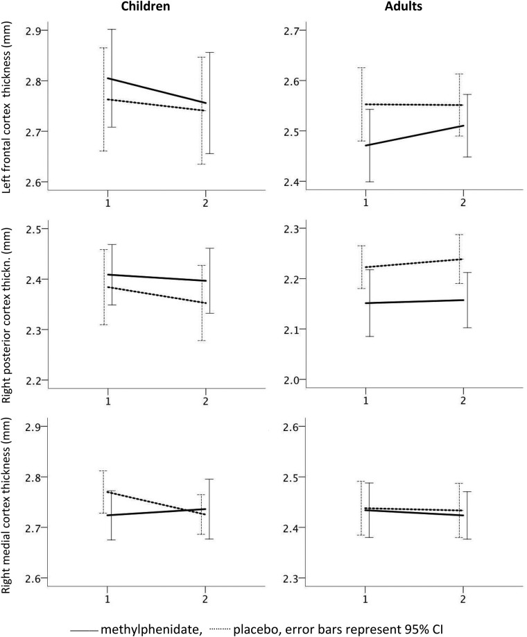
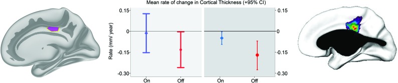
Results: We observed a time × medication × age interaction (F[1,88.825] = 4.316, P < .05) for the right medial cortex ROI, where methylphenidate treatment yielded less cortical thinning in children, but not in adults or the placebo groups.

Conclusions: Our finding that the effects of methylphenidate on right medial cortical thickness differ between children and adults infers that the drug affects gray matter development in this brain region. This warrants replication in larger groups with longer follow-up to determine whether this effect can also be observed in other cortical brain regions and whether it may have long-term consequences.


Figures

Fig 1.
Fig 1.
ROIs investigated. Cortical ROIs were determined on the basis of the MNI coordinates of the vertices corresponding to peak group differences in the observational prospective study of psychostimulant treatment and the developing cortex by Shaw et al. Labels were created at the corresponding vertices on the FreeSurfer average surface. The labels were dilated 15 times, resulting in hexagon-shaped ROIs, covering approximately 550 mm2 when transformed to the individual participants’ brain surfaces. Shown here are the following: 1) the left frontal cortex ROI in lateral (A) and frontal (B) view; 2) the right medial cortex ROI in the medial view; and 3) the right posterior cortex ROI in the lateral view.
Fig 2.
Fig 2.
Consolidated standards of reporting trials flow diagram. Patients were randomized to either methylphenidate or placebo.
Fig 3.
Fig 3.
Differences in rates of cortical thickness change of the ROIs in children and adults taking methylphenidate or placebo medication. For each ROI, at the y-axis, cortical thickness in millimeters (in left frontal, right posterior, and right medial cortices) is shown at baseline (x-axis time point 1) and at the end point (x-axis time point 2) of the trial. Note that as cortical thickness differs across regions and age groups, different origin values for cortical thickness are given across panels to enable visualization, but the scale is otherwise the same, and increments on the y-axes invariably represent 0.1 mm. Mean rates of change are shown with dotted lines for the placebo group and solid lines for the methylphenidate group. Error bars represent the 95% confidence intervals.
Fig 4.
Fig 4.
Mean rate of change in cortical thickness estimated per year for the present RCT and the previous observational study for the right medial cortex region. For children receiving methylphenidate (blue) and placebo (red), cortical change values for the right medial ROI across the duration of the present RCT were converted to an estimated mean rate of change in cortical thickness per year (left panel). This was done for comparison with previous results (right panel) from the observational study from which the ROI was derived (Shaw et al), reporting on the mean rate of cortical change per year across a period of about 4 years.

References

    1. Burcu M, Zito JM, Metcalfe L, et al. Trends in stimulant medication use in commercially insured youths and adults, 2010–2014. JAMA Psychiatry 2016;73:992–93 10.1001/jamapsychiatry.2016.1182 - DOI - PubMed
    1. Trip AM, Visser ST, Kalverdijk LJ, et al. Large increase of the use of psycho-stimulants among youth in the Netherlands between 1996 and 2006. Br J Clin Pharmacol 2009;67:466–68 10.1111/j.1365-2125.2009.03373.x - DOI - PMC - PubMed
    1. Group M. National Institute of Mental Health Multimodal Treatment Study of ADHD follow-up: 24-month outcomes of treatment strategies for attention-deficit/hyperactivity disorder. Pediatrics 2004;113:754–61 10.1542/peds.113.4.754 - DOI - PubMed
    1. Swanson J, Greenhill L, Wigal T, et al. Stimulant-related reductions of growth rates in the PATS. J Am Acad Child Adolesc Psychiatry 2006;45:1304–13 10.1097/01.chi.0000235075.25038.5a - DOI - PubMed
    1. Shaw P, De Rossi P, Watson B, et al. Mapping the development of the basal ganglia in children with attention-deficit/hyperactivity disorder. J Am Acad Child Adolesc Psychiatry.2014;53:780–89. e711 10.1016/j.jaac.2014.05.003 - DOI - PMC - PubMed

Publication types

Substances

Associated data