

Radiolabeled cCPE Peptides for SPECT Imaging of Claudin-4 Overexpression in Pancreatic Cancer
- PMID: 32414951
- PMCID: PMC8679629
- DOI: 10.2967/jnumed.120.243113
Radiolabeled cCPE Peptides for SPECT Imaging of Claudin-4 Overexpression in Pancreatic Cancer
Abstract
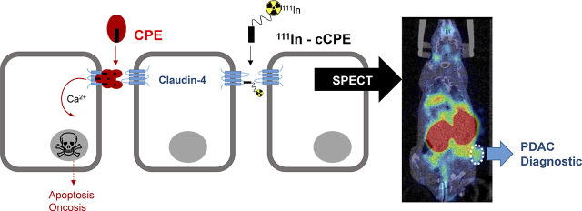
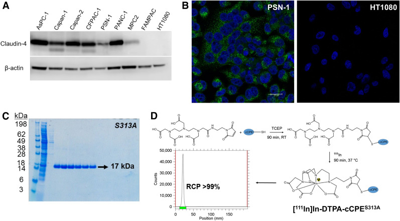
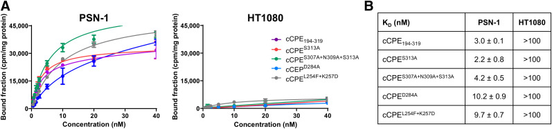
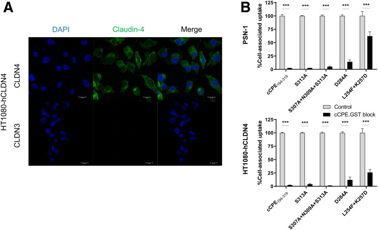
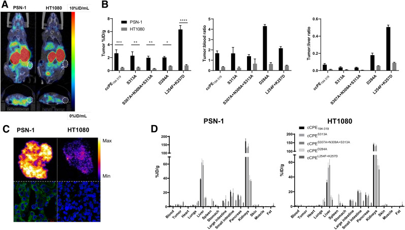
Overexpression of tight-junction protein claudin-4 has been detected in primary and metastatic pancreatic cancer tissue and is associated with better prognosis in patients. Noninvasive measurement of claudin-4 expression by imaging methods could provide a means for accelerating detection and stratifying patients into risk groups. Clostridium perfringens enterotoxin (CPE) is a natural ligand for claudin-4 and holds potential as a targeting vector for molecular imaging of claudin-4 overexpression. A glutathione S-transferases (GST)-tagged version of the C terminus of CPE (cCPE) was previously used to delineate claudin-4 overexpression by SPECT but showed modest binding affinity and slow blood clearance in vivo. Methods: On the basis of the crystal structure of cCPE, a series of smaller cCPE194-319 mutants with putatively improved binding affinity for claudin-4 was generated by site-directed mutagenesis. All peptides were conjugated site-specifically on a C-terminal cysteine using maleimide-diethylenetriamine pentaacetate to enable radiolabeling with 111In. The binding affinity of all radioconjugates was evaluated in claudin-4-expressing PSN-1 cells and HT1080-negative controls. The specificity of all cCPE mutants to claudin-4 was assessed in HT1080 cells stably transfected with claudin-4. SPECT/CT imaging of BALB/c nude mice bearing PSN-1 or HT1080 tumor xenografts was performed to determine the claudin-4-targeting ability of these peptides in vivo. Results: Uptake of all cCPE-based radioconjugates was significantly higher in PSN-1 cells than in HT1080-negative controls. All peptides showed a marked improvement in affinity for claudin-4 in vitro when compared with previously reported values (dissociation constant: 2.2 ± 0.8, 3 ± 0.1, 4.2 ± 0.5, 10 ± 0.9, and 9.7 ± 0.7 nM). Blood clearance of [111In]In-cCPE194-319, as measured by SPECT, was considerably faster than that of [111In]In-cCPE.GST (half-life, <1 min). All radiopeptides showed significantly higher accumulation in PSN-1 xenografts than in HT1080 tumors at 90 min after injection of the tracer ([111In]In-cCPE194-319, 2.7 ± 0.8 vs. 0.4 ± 0.1 percentage injected dose per gram [%ID/g], P < 0.001; [111In]In-S313A, 2.3 ± 0.9 vs. 0.5 ± 0.1 %ID/g, P < 0.01; [111In]In-S307A + N309A + S313A, 2 ± 0.4 vs. 0.3 ± 0.1 %ID/g, P < 0.01; [111In]In-D284A, 2 ± 0.2 vs. 0.7 ± 0.1 %ID/g, P < 0.05; [111In]In-L254F + K257D, 6.3 ± 0.9 vs. 0.7 ± 0.2 %ID/g, P < 0.001). Conclusion: These optimized cCPE-based SPECT imaging agents show great promise as claudin-4-targeting vectors for in vivo imaging of claudin-4 overexpression in pancreatic cancer.
Keywords: SPECT imaging; claudin-4; early diagnosis; pancreatic ductal adenocarcinoma.
© 2020 by the Society of Nuclear Medicine and Molecular Imaging.
Figures
References
-
- GBD 2017 Pancreatic Cancer Collaborators. The global, regional, and national burden of pancreatic cancer and its attributable risk factors in 195 countries and territories, 1990-2017: a systematic analysis for the Global Burden of Disease Study 2017. Lancet Gastroenterol Hepatol. 2019;4:934–947. - PMC - PubMed
-
- SEER cancer statistics review (CSR) 1975-2016. Surveillance, Epidemiology, and End Results Program website. https://seer.cancer.gov/archive/csr/1975_2016/. Updated April 9, 2020. Accessed July 2, 2020.
-
- Ghaneh P, Hanson R, Titman A, et al. . PET-PANC: multicentre prospective diagnostic accuracy and health economic analysis study of the impact of combined modality 18fluorine-2-fluoro-2-deoxy-d-glucose positron emission tomography with computed tomography scanning in the diagnosis and management of pancreatic cancer. Health Technol Assess. 2018;22:1–114. - PMC - PubMed
-
- Higashi T, Saga T, Nakamoto Y, et al. . Diagnosis of pancreatic cancer using fluorine-18 fluorodeoxyglucose positron emission tomography (FDG PET)–usefulness and limitations in “clinical reality.” Ann Nucl Med. 2003;17:261–279. - PubMed
-
- Xu HX, Chen T, Wang WQ, et al. . Metabolic tumour burden assessed by 18F-FDG PET/CT associated with serum CA19-9 predicts pancreatic cancer outcome after resection. Eur J Nucl Med Mol Imaging. 2014;41:1093–1102. - PubMed
Publication types
MeSH terms
Substances
Grants and funding
LinkOut - more resources
Full Text Sources
Medical
Research Materials
Miscellaneous