. 2020 May 15;11(1):2434.
doi: 10.1038/s41467-020-16225-5.

Single cell transcriptomics identifies stem cell-derived graft composition in a model of Parkinson's disease

Affiliations

Single cell transcriptomics identifies stem cell-derived graft composition in a model of Parkinson's disease

Katarína Tiklová et al. Nat Commun. .

Erratum in

Abstract

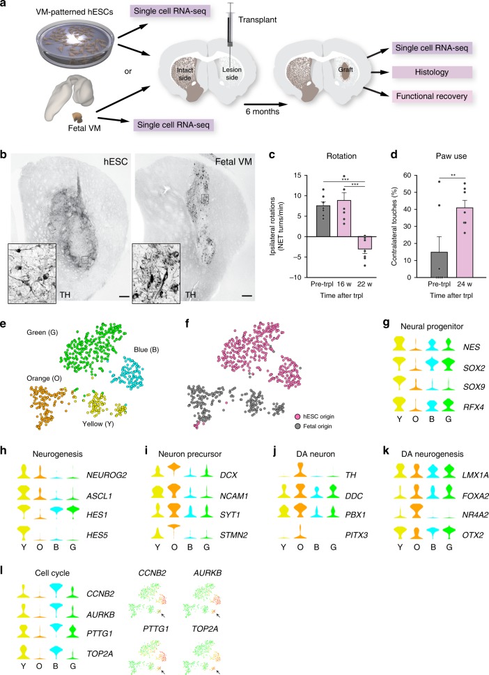
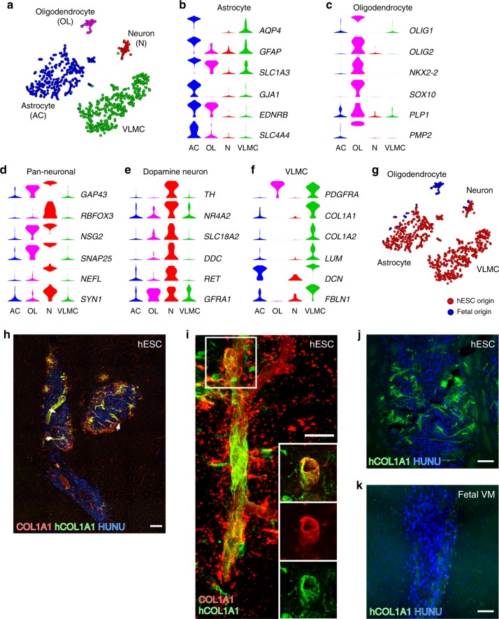
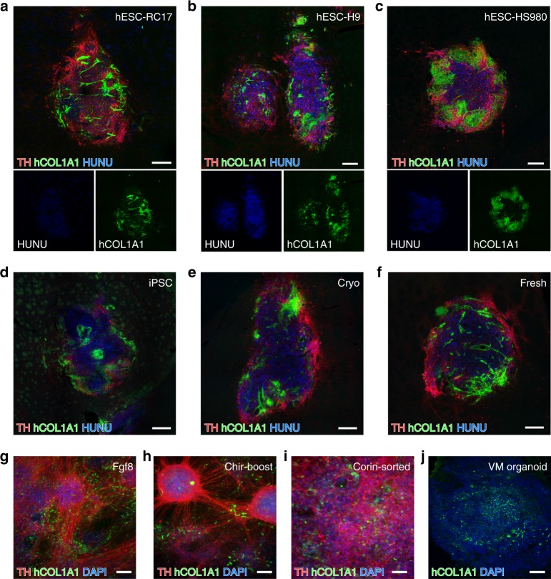
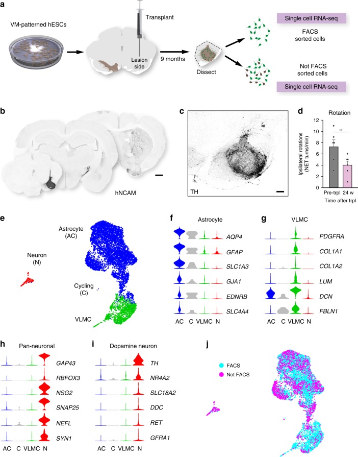
Cell replacement is a long-standing and realistic goal for the treatment of Parkinson's disease (PD). Cells for transplantation can be obtained from fetal brain tissue or from stem cells. However, after transplantation, dopamine (DA) neurons are seen to be a minor component of grafts, and it has remained difficult to determine the identity of other cell types. Here, we report analysis by single-cell RNA sequencing (scRNA-seq) combined with comprehensive histological analyses to characterize intracerebral grafts from human embryonic stem cells (hESCs) and fetal tissue after functional maturation in a pre-clinical rat PD model. We show that neurons and astrocytes are major components in both fetal and stem cell-derived grafts. Additionally, we identify a cell type closely resembling a class of recently identified perivascular-like cells in stem cell-derived grafts. Thus, this study uncovers previously unknown cellular diversity in a clinically relevant cell replacement PD model.


Conflict of interest statement

M.P. is the owner of Parmar Cells AB and co-inventor of the U.S. patent application 15/093,927 owned by Biolamina AB and EP17181588 owned by Miltenyi Biotec. All other authors declare no competing interests.

Figures

Fig. 1
Fig. 1. Histological validation and scRNA-seq analysis of progenitor cells before grafting in a pre-clinical cell therapy model of PD.
a Schematic overview of experimental design. VM-patterned hESCs were grafted to seven rats and used as follows: scRNA-seq n = 2; histology n = 3; functional recovery n = 7. Cells from one fetal VM were grafted to three rats and used as follows: scRNA-seq n = 2; histology n = 1 (n = 1 because of limited access of fetal tissue). b Immunohistochemistry of TH in the graft core of hESC- and fetal VM-derived intrastriatal grafts 6 months post-transplantation. Insets show high-power magnifications of the DA neurons c, d Functional recovery of the hESC-derived cells by amphetamine-induced rotation test and spontaneous paw use (Cylinder) test (n = 7 rats; mean ± SEM; **p < 0.01, ***p < 0.001; compared to post-lesion; two-tailed paired t-test). e t-SNE showing clustering of 660 analyzed cells before grafting (404 cells of hESC origin, 256 cells of fetal origin). Green, blue, orange, and yellow circles define the clusters. f Same t-SNE as in e but with origin of cells marked with pink circles (hESC) or gray circles (fetal) as indicated. gl Expression level per cluster for indicated genes. Genes represent markers for the cell types (neural progenitor, neuron precursor, DA neuron) or indicated processes (cell cycle, neurogenesis, or DA neurogenesis; see text for details). Expression levels of indicated cell cycle genes are also shown in the t-SNE. Scale bar, 250 µM.
Fig. 2
Fig. 2. scRNA-seq analysis and histological validation of grafted cells into the striatum.
a t-SNE showing clustering of 746 cells grafted to striatum (683 cells of hESC origin, grafted rats n = 2; 63 cells of fetal origin, grafted rats n = 2). Cell type assignments are indicated: OL oligodendrocyte, N Neuron, AC astrocyte, VLMC vascular leptomeningeal cells. bd Expression level per cluster for indicated genes. All indicated genes are significantly enriched and established markers for astrocytes, oligodendrocytes, and pan-neuronal cells, respectively. e Expression level per cluster for selected dopaminergic markers. f Expression level per cluster for genes that are significantly enriched in the VLMC cluster and established markers for barrier-forming fibroblasts including VLMCs. g t-SNE of grafted cells as shown in Fig. 2a. Cells are marked according to their origin from either hESC-derived (red circles) or fetal-derived (blue circles) transplants. h Staining using antibodies recognizing both rat and human COL1A1 or only hCOL1A1 as indicated in the core of a hESC-derived graft from the same experiment as used for scRNA-seq. Human nuclei were counterstained with HUNU. i Representative immunofluorescence micrograph of hCOL1A1-positive cells intermingled with host-derived COL1A1-positive cells in close association with blood vessels. Boxed area shows the localization of the close-ups. jk Representative micrographs of hCOL1A1/HUNU immunostaining from a hESC-derived graft (j) and from a fetal-derived grafts from the same experiment as used for scRNA-seq. Scale bars, 200 µM (h), 20 µM (i), and 100 µM (j, k).
Fig. 3
Fig. 3. Presence of VLMCs in different cell lines.
ac Representative immunofluorescence micrograph showing staining of hCOL1A1 in TH-rich hESC-derived grafts from three additional grafting experiments where the transplant was generated from VM-patterned RC17 (a), H9 (b), and HS980 hESC lines (c). d TH/hCOL1A1 immunofluorescence staining of a VM-patterned iPSC-transplant sorted for IAP expression prior to transplantation. e, f TH/hCOL1A1 immunofluorescence staining of VM-patterned hESC-derived grafts generated from cryopreserved cells (e) or fresh cells (f). gi Representative micrographs of TH/hCOL1A1 double immunostaining in terminally differentiated hESC in vitro cultures derived by three different clinically relevant VM-patterning differentiation protocols: the protocol used in this study (g), a protocol developed in the Studer lab that uses CHIR boost instead of FGF8 for proper caudalization (described in https://patents.justia.com/patent/20180094242) (h), and a protocol developed by the Takahashi lab where the cells are sorted based on Corin prior to grafting, (i)). Nuclei were counterstained with DAPI in all three cultures. j hCOL1A immunostaining in a self-organized midbrain patterned organoid. Scale bars, 200 µM (af) and 100 µM (gj).
Fig. 4
Fig. 4. scRNA-seq analysis and histological validation of grafted cells into the midbrain.
a Schematic overview of experimental design. VM-patterned hESCs grafted to midbrain of 6-OHDA rats and analyzed at 9 months (n = 6). These rats were used as follows: scRNA-seq n = 3; histology n = 3, functional recovery n = 6. b Overview of hNCAM fiber outgrowth from hESC-derived intranigral graft showing a neuron-rich graft core and extensive re-innervation of the host striatum. c Immunohistochemistry showing TH staining in graft core of a hESC-derived intranigral graft at 9 months post-transplantation. d Drug-induced rotation test showing functional recovery in rats that have been transplanted to the midbrain with hESC-derived cells (n = 6 rats; mean ± SEM; **p < 0.01; compared to post-lesion; two-tailed paired t-test). e UMAP embedding showing clustering of 7875 analyzed cells after grafting to the midbrain (grafted rats n = 3). fi Expression level per cluster for indicated genes. Indicated genes are established markers for astrocytes, VLMCs, neurons, and DA neurons, respectively. All indicated markers are the same as in Fig. 2. j UMAP of grafted cells as shown in Fig. 4e. Cells isolated by FACS (blue circles, n = 5958) or not by FACS (magenta circles, n = 1917) are indicated. Scale bars, 1 mm (b); 200 µM (c).

Similar articles

Cited by

References

    1. Cooper O, et al. Differentiation of human ES and Parkinson’s disease iPS cells into ventral midbrain dopaminergic neurons requires a high activity form of SHH, FGF8a and specific regionalization by retinoic acid. Mol. Cell Neurosci. 2010;45:258–266. doi: 10.1016/j.mcn.2010.06.017. - DOI - PMC - PubMed
    1. Kriks S, et al. Dopamine neurons derived from human ES cells efficiently engraft in animal models of Parkinson’s disease. Nature. 2011;480:547–551. doi: 10.1038/nature10648. - DOI - PMC - PubMed
    1. Barker RA, Parmar M, Studer L, Takahashi J. Human trials of stem cell-derived dopamine neurons for Parkinson’s disease: dawn of a new era. Stem Cell. 2017;21:569–573. - PubMed
    1. Nolbrant S, Heuer A, Parmar M, Kirkeby A. Generation of high-purity human ventral midbrain dopaminergic progenitors for in vitro maturation and intracerebral transplantation. Nat. Protoc. 2017;12:1962–1979. doi: 10.1038/nprot.2017.078. - DOI - PubMed
    1. Barker, R. A. TRANSEURO consortium. Designing stem-cell-based dopamine cell replacement trials for Parkinson’s disease. Nat. Med.25, 1045–1053 (2019). - PubMed

Publication types

MeSH terms