. 2020 Oct;27(10):2942-2951.
doi: 10.1038/s41418-020-0553-9. Epub 2020 May 15.

Staufen 1 amplifies proapoptotic activation of the unfolded protein response

Affiliations

Staufen 1 amplifies proapoptotic activation of the unfolded protein response

Mandi Gandelman et al. Cell Death Differ. 2020 Oct.

Erratum in

Abstract

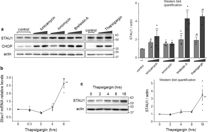
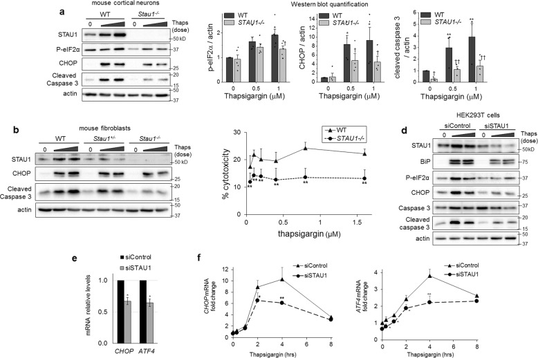
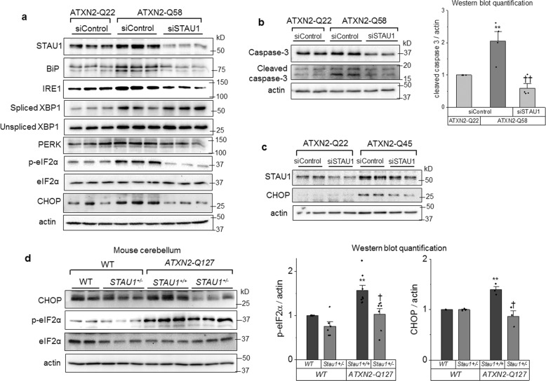
Staufen-1 (STAU1) is an RNA-binding protein that becomes highly overabundant in numerous neurodegenerative disease models, including those carrying mutations in presenilin1 (PSEN1), microtubule-associated protein tau (MAPT), huntingtin (HTT), TAR DNA-binding protein-43 gene (TARDBP), or C9orf72. We previously reported that elevations in STAU1 determine autophagy defects and its knockdown is protective in models of several neurodegenerative diseases. Additional functional consequences of STAU1 overabundance, however, have not been investigated. We studied the role of STAU1 in the chronic activation of the unfolded protein response (UPR), a common feature among neurodegenerative diseases and often directly associated with neuronal death. Here we report that STAU1 is a novel modulator of the UPR, and is required for apoptosis induced by activation of the PERK-CHOP pathway. STAU1 levels increased in response to multiple endoplasmic reticulum (ER) stressors, and exogenous expression of STAU1 was sufficient to cause apoptosis through the PERK-CHOP pathway of the UPR. Cortical neurons and skin fibroblasts derived from Stau1-/- mice showed reduced UPR and apoptosis when challenged with thapsigargin. In fibroblasts from individuals with SCA2 or with ALS-causing TDP-43 and C9ORF72 mutations, we found highly increased STAU1 and CHOP levels in basal conditions, and STAU1 knockdown restored CHOP levels to normal. Taken together, these results show that STAU1 overabundance reduces cellular resistance to ER stress and precipitates apoptosis.


Conflict of interest statement

The authors declare that they have no conflict of interest.

Figures

Fig. 1
Fig. 1. STAU1 overabundance induced by ER stress or calcium dyshomeostasis.
a HEK293 cells were incubated with tunicamycin (0.1 and 0.5 µM), ionomycin (0.5 and 1 µM), brefeldin A (0.5 and 1 µM), and thapsigargin (0.5 and 1 µM) for 18 h. Levels of STAU1 and CHOP were evaluated by western blot. Graph represents quantification of STAU1/actin from three independent experiments. Single asterisk (*) or double asterisks (**) denote significantly different from untreated control. b Relative STAU1 mRNA levels in HEK293 cells treated with thapsigargin (0.5 µM) for the times indicated. c Levels of STAU1 protein in HEK293 cells treated with thapsigargin (0.5 µM) for the times indicated. Graph represents quantification of STAU1/actin for three independent experiments. Data are mean ± SEM. *p < 0.05 and **p < 0.01 according to the paired sample t test.
Fig. 2
Fig. 2. Attenuated UPR and apoptosis in cells deficient in STAU1.
Western blots of cultured cortical neurons (a) or skin fibroblasts (b) from WT, Stau1+/−, or Stau1−/− mice incubated with thapsigargin (0.25 and 0.5 µM, 18 h). Graph represents quantification of target protein/actin from three independent experiments. c Cytotoxicity assessment by quantification of LDH release in WT or Stau1−/− mouse fibroblasts exposed to indicated doses of thapsigargin for 24 h. d Western blots of HEK293 cells transfected with siControl or siSTAU1 for 72 h and incubated with thapsigargin (0.5 and 1 µM) for 18 h. e mRNA levels of CHOP and ATF4 in HEK293 72 h post transfection with siControl or siSTAU1 and f after treatment with thapsigargin (0.5 µM) for the times indicated. Single asterisk (*), double asterisks (**), single dagger (), or double daggers (††) denote significantly different from WT or siControl treated with the corresponding dose of thapsigargin. Data are mean ± SEM. * or p < 0.05, ** or ††p < 0.01 by two-way ANOVA.
Fig. 3
Fig. 3. Exogenous STAU1 induces apoptosis through the PERK pathway of the UPR.
HEK293 cells were transfected with 3xFlag-STAU1 (3xF-STAU1) or empty vector control (3xF) with addition of siRNA directed at PERK (siPERK) after 24 h or the PERK inhibitor GSK2606414 (0.5 µM) after 48 h. After a further 18 h, protein levels were analyzed by western blot. Graphs represent the quantification of three independent experiments. Single asterisk (*) or double asterisks (**) denote significantly different from 3xF control. Single dagger () or double daggers (††) denote significantly different from same genotype control. Data are mean ± SEM. ∗ or p < 0.05, ∗∗ or ††p < 0.01 according to the paired sample t test.
Fig. 4
Fig. 4. Attenuation of UPR and apoptosis in cellular and animal models of SCA2 by siSTAU1 or genetic interaction.
a Western blot of proteins involved in the UPR, and b caspase 3 and cleaved caspase 3 in ATXN2-Q22 and ATXN2-Q58 cells. Graph represents quantification of three independent experiments. c Western blot of fibroblasts derived from an SCA2 patient with ATXN2-Q45 mutation. d Western blot of cerebellar tissue from WT, ATXN2-Q127 mice and Stau1+/− haploinsufficient littermates at 34 weeks of age. Graph represents quantification of three animals per genotype. Single asterisk (*) or double asterisks (**) denote significantly different from ATXN2-Q22 or WT control. Single dagger () or double daggers (††) denote significantly different from same genotype control. Data are mean ± SEM. * or p < 0.05, ** or ††p < 0.01 according to the paired sample t test.
Fig. 5
Fig. 5. STAU1 knockdown reduces the proapoptotic activation of the UPR in fibroblasts from patients with ALS and FTD-causing mutations.
a Western blot of STAU1, p-eIF2α, and CHOP in fibroblasts derived from human subjects without disease-related mutation (normal), with TARDBP A382T (line 1) and one with TARDBP G298S (line 2) (b) and two individuals with C9ORF72 GGGGCC repeat expansion (lines 1 and 2). Single asterisk (*) or double asterisks (**) denote significantly different from control patient. Single dagger () or double daggers (††) denote significantly different from same genotype control. Data are mean ± SEM. * or p < 0.05, ** or ††p < 0.01 according to the paired sample t test.

References

    1. Paul S, Dansithong W, Gandelman M, Zu T, Ranum LPW, Figueroa KP, et al. Staufen blocks autophagy in neurodegeneration. 10.1101/659649. 2019.
    1. Paul S, Dansithong W, Figueroa KP, Scoles DR, Pulst SM. Staufen1 links RNA stress granules and autophagy in a model of neurodegeneration. Nat Commun. 2018;9:3648. doi: 10.1038/s41467-018-06041-3. - DOI - PMC - PubMed
    1. Liu J, Zhang KS, Hu B, Li SG, Li Q, Luo YP, et al. Systematic analysis of RNA regulatory network in rat brain after ischemic stroke. Biomed Res Int. 2018;2018:8354350. - PMC - PubMed
    1. Krichevsky AM, Kosik KS. Neuronal RNA granules: a link between RNA localization and stimulation-dependent translation. Neuron. 2001;32:683–96. doi: 10.1016/S0896-6273(01)00508-6. - DOI - PubMed
    1. Vessey JP, Macchi P, Stein JM, Mikl M, Hawker KN, Vogelsang P, et al. A loss of function allele for murine Staufen1 leads to impairment of dendritic Staufen1-RNP delivery and dendritic spine morphogenesis. Proc Natl Acad Sci USA. 2008;105:16374–9. doi: 10.1073/pnas.0804583105. - DOI - PMC - PubMed

Publication types