

Branch-type intraductal papillary neoplasm of the bile duct treated with laparoscopic anatomical resection: a case report
- PMID: 32415464
- PMCID: PMC7229076
- DOI: 10.1186/s40792-020-00864-3
Branch-type intraductal papillary neoplasm of the bile duct treated with laparoscopic anatomical resection: a case report
Abstract
Background: Intraductal papillary neoplasm of the bile duct (IPNB) is characterized by an intraluminal, growing papillary tumor covered by neoplastic biliary epithelial cells with a fine fibrovascular core. IPNB was introduced as a precancerous and early neoplastic lesion in the 2010 World Health Organization classification of tumors of the digestive system. IPNB eventually invades the bile duct wall and progresses to invasive cholangiocarcinoma. IPNB resembles intraductal papillary mucinous neoplasm of the pancreas (IPMN), particularly the main pancreatic duct type. IPNB cases, possibly corresponding to branch-type IPMN, have been recently reported, and these cases involved the peribiliary glands significantly and showed gross cystic dilatation. Small branch-type intrahepatic IPNB often mimics simple liver cysts, making the diagnosis of IPNB difficult. Some literature recommended surgical resection for treatment. Laparoscopic resection is a good treatment option for small tumor. We herein present the case of branch-type IPNB that was treated with laparoscopic anatomical liver resection 5 years after being detected.
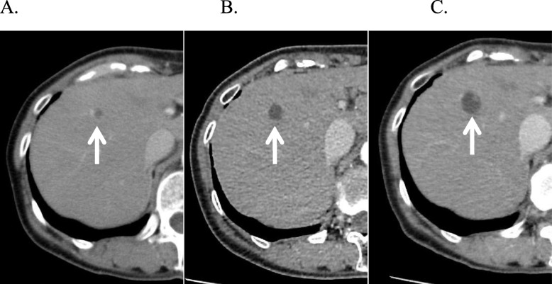
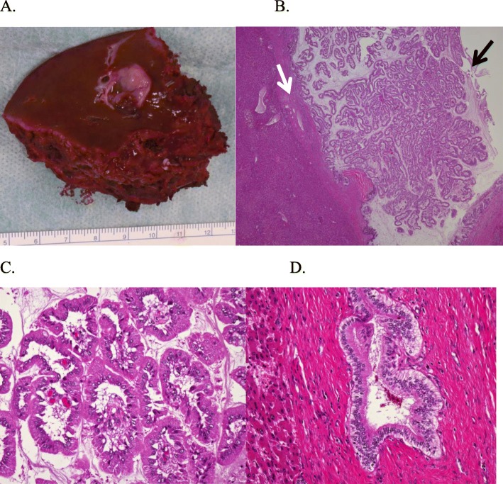
Case presentation: A 64-year-old woman was undergoing follow-up for primary aldosteronism. In 2012, follow-up computed tomography (CT) incidentally revealed a 7-mm cystic lesion in segment 8 of the liver. From 2012 to 2017, the cystic lesion kept increasing in size, reaching 17 mm. In 2017, CT also revealed a 13-mm mural nodule in the cyst wall. Therefore, the patient was referred to our department for possible malignancy. We suspected a branch-type IPNB; however, the mass was small and diagnosis could not be made without performing biopsy. Accordingly, surgical resection was performed for diagnosis and treatment. Because branch-type IPNB might show horizontal spread through the intrahepatic bile duct, we believed that anatomical resection of the liver was appropriate considering the malignant potential of the lesion. Therefore, laparoscopic anatomical resection of segment 8 of the liver was performed. The resected tumor measured 17 mm and was histologically diagnosed as a high-grade IPNB.
Conclusion: Branch-type IPNBs are rare but can potentially lead to malignant tumors. Surgical resection is the treatment of choice, with laparoscopic anatomical resection being a good treatment option for this small tumor.
Keywords: Intraductal papillary mucinous neoplasm of the pancreas; Intraductal papillary neoplasm of the bile duct; Laparoscopic anatomical resection; Segmentectomy; Surgical margin.
Conflict of interest statement
The authors declare that they have no competing interests.
Figures


Similar articles
-
Intraductal Papillary Neoplasm of Bile Duct: Updated Clinicopathological Characteristics and Molecular and Genetic Alterations.J Clin Med. 2020 Dec 9;9(12):3991. doi: 10.3390/jcm9123991. J Clin Med. 2020. PMID: 33317146 Free PMC article. Review.
-
Recurrent intraductal papillary neoplasm of the bile duct due to intraductal dissemination: a case report and literature review.Surg Case Rep. 2021 Nov 5;7(1):238. doi: 10.1186/s40792-021-01318-0. Surg Case Rep. 2021. PMID: 34739634 Free PMC article.
-
Lung metastases from intraductal papillary neoplasm of the bile duct: a case report.World J Surg Oncol. 2020 Oct 23;18(1):271. doi: 10.1186/s12957-020-02054-9. World J Surg Oncol. 2020. PMID: 33097064 Free PMC article.
-
Intraductal Papillary Mucinous Neoplasm of the Bile Duct: A Case Report.Cureus. 2025 Feb 8;17(2):e78749. doi: 10.7759/cureus.78749. eCollection 2025 Feb. Cureus. 2025. PMID: 40070623 Free PMC article.
-
A statement by the Japan-Korea expert pathologists for future clinicopathological and molecular analyses toward consensus building of intraductal papillary neoplasm of the bile duct through several opinions at the present stage.J Hepatobiliary Pancreat Sci. 2018 Mar;25(3):181-187. doi: 10.1002/jhbp.532. Epub 2018 Feb 12. J Hepatobiliary Pancreat Sci. 2018. PMID: 29272078 Review.
Cited by
-
Intrahepatic intraductal papillary cystic neoplasm of the bile duct: A case report.Ann Med Surg (Lond). 2021 Feb 12;63:102167. doi: 10.1016/j.amsu.2021.02.013. eCollection 2021 Mar. Ann Med Surg (Lond). 2021. PMID: 33664950 Free PMC article.
-
Diagnosing rare intraductal biliary neoplasms - Intraductal papillary neoplasm of the bile duct: A case report with typical imaging findings.SA J Radiol. 2022 Apr 29;26(1):2387. doi: 10.4102/sajr.v26i1.2387. eCollection 2022. SA J Radiol. 2022. PMID: 35548709 Free PMC article.
-
Intraductal Papillary Neoplasm of the Bile Duct: A Rare Disease and Presentation.Cureus. 2023 Feb 2;15(2):e34556. doi: 10.7759/cureus.34556. eCollection 2023 Feb. Cureus. 2023. PMID: 36879718 Free PMC article.
-
Clinical characteristics of intrahepatic biliary papilloma: A case report.World J Clin Cases. 2021 May 6;9(13):3185-3193. doi: 10.12998/wjcc.v9.i13.3185. World J Clin Cases. 2021. PMID: 33969107 Free PMC article.
-
Intraductal Papillary Neoplasm of Bile Duct: Updated Clinicopathological Characteristics and Molecular and Genetic Alterations.J Clin Med. 2020 Dec 9;9(12):3991. doi: 10.3390/jcm9123991. J Clin Med. 2020. PMID: 33317146 Free PMC article. Review.
References
-
- Nakamura Y, Curabo MO, Franceschi S, et al. Intrahepatic cholangiocarcinoma. WHO Classification of Tumors of the Digestive System World Health Organization of Tumors, 4th edn. LYON: IARC 2010;217-224.
LinkOut - more resources
Full Text Sources
