

Lipid Rafts from Olfactory Ensheathing Cells: Molecular Composition and Possible Roles
- PMID: 32415577
- PMCID: PMC11448638
- DOI: 10.1007/s10571-020-00869-4
Lipid Rafts from Olfactory Ensheathing Cells: Molecular Composition and Possible Roles
Abstract
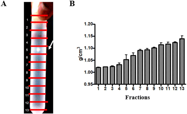
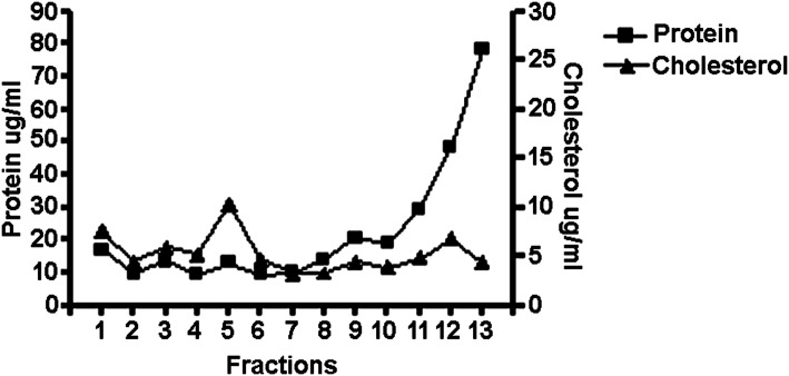
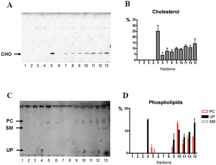
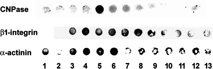
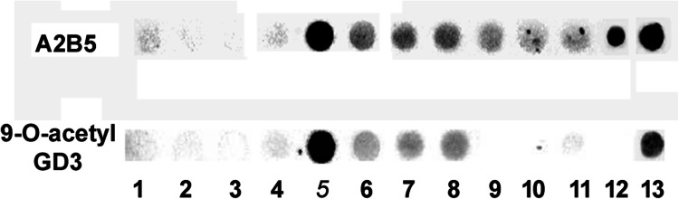
Olfactory ensheathing cells (OECs) are specialized glial cells of the olfactory system, believed to play a role in the continuous production of olfactory neurons and ensheathment of their axons. Although OECs are used in therapeutic applications, little is known about the cellular mechanisms underlying their migratory behavior. Recently, we showed that OEC migration is sensitive to ganglioside blockage through A2B5 and Jones antibody in OEC culture. Gangliosides are common components of lipid rafts, where they participate in several cellular mechanisms, including cell migration. Here, we characterized OEC lipid rafts, analyzing the presence of specific proteins and gangliosides that are commonly expressed in motile neural cells, such as young neurons, oligodendrocyte progenitors, and glioma cells. Our results showed that lipid rafts isolated from OECs were enriched in cholesterol, sphingolipids, phosphatidylcholine, caveolin-1, flotillin-1, gangliosides GM1 and 9-O-acetyl GD3, A2B5-recognized gangliosides, CNPase, α-actinin, and β1-integrin. Analysis of the actin cytoskeleton of OECs revealed stress fibers, membrane spikes, ruffled membranes and lamellipodia during cell migration, as well as the distribution of α-actinin in membrane projections. This is the first description of α-actinin and flotillin-1 in lipid rafts isolated from OECs and suggests that, together with β1-integrin and gangliosides, membrane lipid rafts play a role during OEC migration. This study provides new information on the molecular composition of OEC membrane microdomains that can impact on our understanding of the role of OEC lipid rafts under physiological and pathological conditions of the nervous system, including inflammation, hypoxia, aging, neurodegenerative diseases, head trauma, brain tumor, and infection.
Keywords: Cell motility; Gangliosides; Glial cells; Membrane microdomains.
Conflict of interest statement
Authors declare that they have no conflict of interest.
Figures


Similar articles
-
A role for gangliosides and β1-integrin in the motility of olfactory ensheathing glia.J Anat. 2019 Nov;235(5):977-983. doi: 10.1111/joa.13057. Epub 2019 Aug 2. J Anat. 2019. PMID: 31373393 Free PMC article.
-
Defining the morphological phenotype: 2',3'-cyclic nucleotide 3'-phosphodiesterase (CNPase) is a novel marker for in situ detection of canine but not rat olfactory ensheathing cells.Cell Tissue Res. 2011 Jun;344(3):391-405. doi: 10.1007/s00441-011-1168-8. Epub 2011 Apr 26. Cell Tissue Res. 2011. PMID: 21519895
-
Streptococcus pneumoniae infection regulates expression of neurotrophic factors in the olfactory bulb and cultured olfactory ensheathing cells.Neuroscience. 2016 Mar 11;317:149-61. doi: 10.1016/j.neuroscience.2016.01.016. Epub 2016 Jan 12. Neuroscience. 2016. PMID: 26791522
-
Interaction of membrane/lipid rafts with the cytoskeleton: impact on signaling and function: membrane/lipid rafts, mediators of cytoskeletal arrangement and cell signaling.Biochim Biophys Acta. 2014 Feb;1838(2):532-45. doi: 10.1016/j.bbamem.2013.07.018. Epub 2013 Jul 27. Biochim Biophys Acta. 2014. PMID: 23899502 Free PMC article. Review.
-
Lipid rafts and neurodegeneration: structural and functional roles in physiologic aging and neurodegenerative diseases.J Lipid Res. 2020 May;61(5):636-654. doi: 10.1194/jlr.TR119000427. Epub 2019 Dec 23. J Lipid Res. 2020. PMID: 31871065 Free PMC article. Review.
Cited by
-
Interactive mechanisms between caveolin-1 and actin filaments or vimentin intermediate filaments instruct cell mechanosensing and migration.J Mol Cell Biol. 2023 Apr 6;14(11):mjac066. doi: 10.1093/jmcb/mjac066. J Mol Cell Biol. 2023. PMID: 36472547 Free PMC article. No abstract available.
-
Olfactory ensheathing cells and neuropathic pain.Front Cell Dev Biol. 2023 Apr 5;11:1147242. doi: 10.3389/fcell.2023.1147242. eCollection 2023. Front Cell Dev Biol. 2023. PMID: 37223000 Free PMC article. Review.
-
9-O Acetylated Gangliosides in Health and Disease.Biomolecules. 2023 May 12;13(5):827. doi: 10.3390/biom13050827. Biomolecules. 2023. PMID: 37238697 Free PMC article. Review.
-
Multi-Omics Prognostic Signatures Based on Lipid Metabolism for Colorectal Cancer.Front Cell Dev Biol. 2022 Feb 11;9:811957. doi: 10.3389/fcell.2021.811957. eCollection 2021. Front Cell Dev Biol. 2022. PMID: 35223868 Free PMC article.
-
Elucidating the Pivotal Neuroimmunomodulation of Stem Cells in Spinal Cord Injury Repair.Stem Cells Int. 2021 Jul 23;2021:9230866. doi: 10.1155/2021/9230866. eCollection 2021. Stem Cells Int. 2021. PMID: 34341666 Free PMC article. Review.
References
-
- Bligh EG, Dyer WJ (1959) A rapid method of total lipid extraction and purification. Can J Biochem Physiol 37:911–913 - PubMed
-
- Brown DA (2002) Isolation and use of rafts. Curr Protoc Immunol 51:11 - PubMed
-
- Corrêa JR, Atella GC, Vargas C, Soares MJ (2007) Transferrin uptake may occur through detergent-resistant membrane domains at the cytopharynx of Trypanosoma cruzi epimastigote forms. Mem Inst Oswaldo Cruz 102:871–876 - PubMed
-
- Del Pozo MA (2004) Integrin signaling and lipid rafts. Cell Cycle 3:725–728 - PubMed
MeSH terms
Substances
LinkOut - more resources
Full Text Sources
