Review
. 2020:165-166:142-154.
doi: 10.1016/j.addr.2020.05.004. Epub 2020 May 13.

Orally ingestible medical devices for gut engineering

Affiliations
Review

Orally ingestible medical devices for gut engineering

Nikolaj Kofoed Mandsberg et al. Adv Drug Deliv Rev. 2020.

Abstract

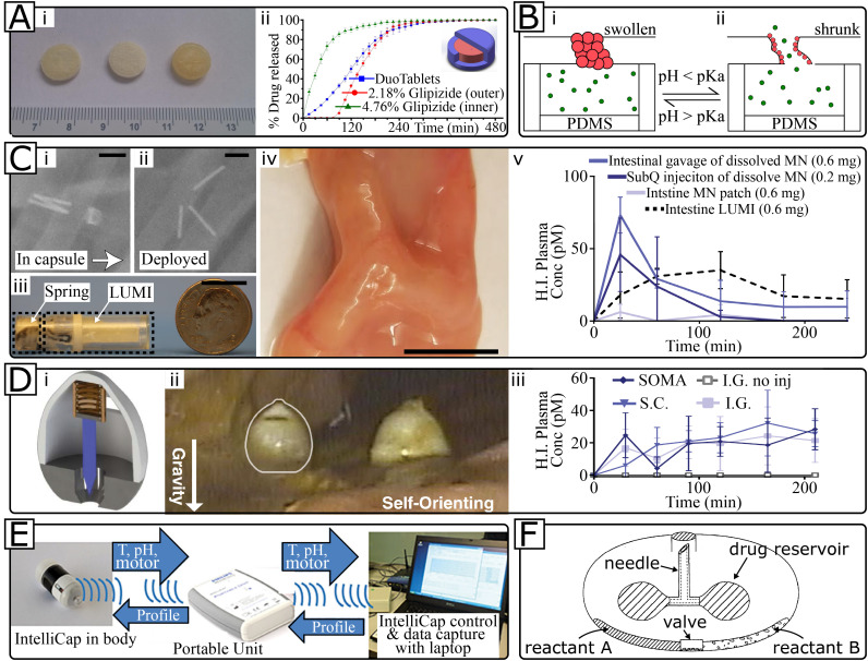
Orally ingestible medical devices provide significant advancement for diagnosis and treatment of gastrointestinal (GI) tract-related conditions. From micro- to macroscale devices, with designs ranging from very simple to complex, these medical devices can be used for site-directed drug delivery in the GI tract, real-time imaging and sensing of gut biomarkers. Equipped with uni-direction release, or self-propulsion, or origami design, these microdevices are breaking the barriers associated with drug delivery, including biologics, across the GI tract. Further, on-board microelectronics allow imaging and sensing of gut tissue and biomarkers, providing a more comprehensive understanding of underlying pathophysiological conditions. We provide an overview of recent advances in orally ingestible medical devices towards drug delivery, imaging and sensing. Challenges associated with gut microenvironment, together with various activation/actuation modalities of medical devices for micromanipulation of the gut are discussed. We have critically examined the relationship between materials-device design-pharmacological responses with respect to existing regulatory guidelines and provided a clear roadmap for the future.

Keywords: GI diagnostics; Gastrointestinal tract; Gut microbiota; Medical microdevices; Oral drug delivery.


Figures

Unlabelled Image
Graphical abstract
Fig. 1
Fig. 1
Orally ingestible microdevices: 1A) Timeline depicting the first appearance of multi-compartment sensing, drug delivery, and sampling devices to illustrate technological progression and the focus of our review; 1B) Size scale as a selection criteria for oral device application in sensing, drug delivery and gut microsampling.
Fig. 2
Fig. 2
Schematic of the gastrointestinal (GI) tract with site-specific pH ranges, average transit times and predominant enzymes [[24], [25], [26]]. Additional challenges faced by orally ingested microdevices traversing through the GI tract have been highlighted. Images adapted from Servier Medical Art by Servier and licensed under a Creative Commons Attribution 3.0 Unported License.
Fig. 3
Fig. 3
Examples of passive and active microdevices for oral drug delivery and as advanced in vitro cell model. 3A) Pharmacokinetic profile showing the enhanced in vivo bioavailability of acyclovir using planar microdevices (inset) compared to an oral solution with the same concentration of acyclovir. Reprinted from [30] with permission. 3B) Schematic of the printing process of bottom-up fabricated enteric devices for oral delivery. The polymer dispersion was ejected onto the silicon wafer by a picoliter dispenser, where after evaporation of the solvent forms the device body. The same dispenser was used to print drug formulation into each device and a second polymer was applied on top to seal the device before removal from the silicon wafer. Adapted from [36] with permission. 3C) Human gut-on-a-chip microdevice. (i) Photograph of the microdevice, where blue and red dyes fill the upper and lower channels, respectively. (ii) Cross-sectional schematic of the device showing how suction to side channels (grey arrows) applies peristalsis-like mechanical constrictions and fluid flow (white arrows) generates shear stress. (iii) Micrograph showing intestinal basal crypt (red arrow) and villi (white arrow) formed by human Caco-2 cells grown for ~100 h in the microdevice. (iv) Confocal immunofluorescence image showing a horizontal cross-section of intestinal villi similar to the ones shown in (iii). Scale bars represent 50 μm unless indicated otherwise on Fig. 3C. Reprinted from [43] with permission. 3D) Self-propelled microrockets for targeted drug delivery in the stomach. (i) SEM image of a full DOX/poly (aspartic acid)/Fe-Zn microrocket and (ii) energydispersive X-ray spectroscopy mappings of Zn inside the microrocket. (iii) Superimposed fluorescent images of the whole stomachs of mice collected 30 min after administration of ultrapure water and (iv) DOX/poly (aspartic acid)/Pt microrockets and (v) DOX/poly (aspartic acid)/Fe-Zn microrockets. (vi) Histological evaluation of gastric tissue 24 h after administration of poly (aspartic acid)/Fe-Zn microrockets and (vii) water. Reprinted with permission from Zhou et al., Self-propelled and targeted drug delivery of poly(aspartic acid)/iron-zinc microrocket in the stomach, ACS Nano, 13. Copyright 2019 American Chemical Society. 3E) Biomimetic micromotors for delivery of antigens for oral vaccination. (i) Schematic of the formulation concept; after oral ingestion of micromotors, the coating is dissolved in the small intestine, which activates the motor to provide enhanced retention and stimulation. (ii) Images of the GI tract of mice 6 h after oral administration of labeled static microparticles or (iii) micromotors. (iv) Data showing a significantly higher level of IgA titers against α-toxin after administration of micromotors compared to static microparticles. Reprinted with permission from Wei et al., Biomimetic micromotor enables active delivery of antigens for oral vaccination, Nano Letters, 19. Copyright 2019 American Chemical Society. 3F) Micromotors for local treatment of stomach infection in vivo. (i) Schematic of the preparation of micromotors; a dispersion of Mg microparticles is dispersed on a glass slide followed by TiO2 atomic layer deposition and coating with drug-loaded PLGA and chitosan. (ii) Time-lapse images of the propulsion of the micromotors after 2, 4 and 6 min in simulated gastric fluid (pH ~1.3). (iii) Retention of the micromotors visualized with bright-field and fluorescence overlay images of freshly removed mouse stomachs 0 h after oral gavage of ultrapure water as control and 2 h after oral gavage of micromotors. Shared under a Creative Commons Attribution 4.0 International License with copyrights reserved with the authors [51]. (For interpretation of the references to colour in this figure legend, the reader is referred to the web version of this article.)
Fig. 4
Fig. 4
Examples of orally ingested macrodevices for drug delivery. 4A) 3D-printed DuoTablet with controllable release characteristics. (i) Photograph of three such tablets and (ii) their respective release profiles. Reprinted from International Journal of Pharmaceutics, 525 (1), Li et al., Preparation and investigation of controlled-release glipizide novel oral device with three-dimensional printing, 5–11, Copyright 2017, with permission from Elsevier. 4B) Polymeric PNIPAm-MAA nanoparticles shrink upon pH decrease, thereby opening a channel for releasing the drug contained. Schematics shown of (i) the high pH swollen/closed state and (ii) the low pH shrunk/open state. 4C) Luminal Unfolding Microneedle Injection (LUMI) device (scale bars: 1 cm). Swine in vivo radiographs showing (i) the encapsulated/folded state and (ii) the deployed/unfolded state. (iii) Photograph of the encapsulated LUMI and (iv) deployed inside a small intestine to show needle-to-wall contact regardless of device orientation. (v) In vivo human insulin delivery in swine by four different methods. Reprinted by permission from Springer Nature: Nature Medicine, A luminal unfolding microneedle injector for oral delivery of macromolecules, Abramson et al., Copyright 2019. 4D) Self-Orienting Millimeter-scale Applicator (SOMA). (i) A sketch of the device showing the initially compressed spring that will force insertion of the drug-loaded millipost. (ii) The devices have weighted metal bottoms that self-orients in the stomach, which shows here with in vivo endoscopy in fasted swine. (iii) Blood plasma levels of human insulin delivered by four different methods to swine in vivo model. From [[73]]. Reprinted with permission from AAAS. 4E) IntelliCap® is a device for controlled release drug delivery, which can be pre-programmed, real-time controlled, or triggered at specific temperature/pH changes to deliver with different rates. 4F) Balloon based delivery device. Both the needle injection and liquid drug pumping are actuated by a balloon being inflated upon mixing of reactant A and B.
Fig. 5
Fig. 5
Examples of orally ingested macrodevices for sensing and sampling. 5A) The first wireless capsule endoscope: PillCam. (i) The endoscope features two wide-angle cameras and a transmitter for real-time monitoring on (ii) Data Recorder 3. 5B) Ingestible Micro-Bio-Electronic Device (IMBED) uses genetically engineered bacteria to detect the presence of biomarkers. (i) Schematic showing the working principle with light generating bacteria and photodectors. (ii) Diagram of the electronic processing: all the way from detecting the light and wirelessly transmitting the information to the operating personnel. (iii) X-ray and (iv) endoscopy images illustrating the location of the device in a swine stomach (scale bar: 5 cm). (v) The generated photocurrent during an intestinal bleeding detection (blood) and a reference (buffer) experiment. From [[91]]. Reprinted with permission from AAAS. 5C) Mechanical suction device for sampling of GI material. A spring is relaxed upon dissolution of a compressing material, thereby creating a partial vacuum that opens the valves and draws in GI fluids. 5D) Osmotic pressure driven suction device for continuous sampling of intestinal material. (i) Overview of the device showing channel system and the osmotic-pressure-creating semipermeable membrane with salt chamber. (ii) The device has an imbedded magnet for optional prolonged retention in locations of extra interest and (iii) can sample continuously for 48 h with near-constant rate. (iv) The device can sample in both pH 4 and 8 environments, but with different rates. (v) The collected bacteria from the microbiome continuous to multiple within the device after their sampling.

References

    1. Learmonth I.D., Young C., Rorabeck C. The operation of the century: Total hip replacement. Lancet. 2007 doi: 10.1016/S0140-6736(07)60457-7. - DOI - PubMed
    1. Wilson B.S., Finley C.C., Lawson D.T., Wolford R.D., Eddington D.K., Rabinowitz W.M. Better speech recognition with Cochlear implants. Nature. 1991 doi: 10.1038/352236a0. - DOI - PubMed
    1. Otsuka F., Vorpahl M., Nakano M., Foerst J., Newell J.B., Sakakura K., Kutys R., Ladich E., Finn A.V., Kolodgie F.D. Pathology of second-generation Everolimus-eluting stents versus first-generation Sirolimus-and paclitaxel-eluting stents in humans. Circulation. 2014 doi: 10.1161/CIRCULATIONAHA.113.001790. - DOI - PMC - PubMed
    1. Nelson G.D. A brief history of cardiac pacing. Tex. Heart Inst. J. 1993;20(1):12–18. - PMC - PubMed
    1. Golub J.S., Ling L., Nie K., Nowack A., Shepherd S.J., Bierer S.M., Jameyson E., Kaneko C.R.S., Phillips J.O., Rubinstein J.T. Prosthetic implantation of the human vestibular system. Otol. Neurotol. 2014 doi: 10.1097/MAO.0000000000000003. - DOI - PMC - PubMed

Publication types

LinkOut - more resources