. 2020 Nov;223(5):733.e1-733.e14.
doi: 10.1016/j.ajog.2020.05.012. Epub 2020 May 15.

Simvastatin ameliorates altered mechanotransduction in uterine leiomyoma cells

Affiliations

Simvastatin ameliorates altered mechanotransduction in uterine leiomyoma cells

Sadia Afrin et al. Am J Obstet Gynecol. 2020 Nov.

Abstract

Background: Uterine leiomyomas, the most common tumors of the female reproductive system, are characterized by excessive deposition of disordered stiff extracellular matrix and fundamental alteration in the mechanical signaling pathways. Specifically, these alterations affect the normal dynamic state of responsiveness to mechanical cues in the extracellular environment. These mechanical cues are converted through integrins, cell membrane receptors, to biochemical signals including cytoskeletal signaling pathways to maintain mechanical homeostasis. Leiomyoma cells overexpress β1 integrin and other downstream mechanical signaling proteins. We previously reported that simvastatin, an antihyperlipidemic drug, has antileiomyoma effects through cellular, animal model, and epidemiologic studies.

Objective: This study aimed to examine the hypothesis that simvastatin might influence altered mechanotransduction in leiomyoma cells.

Study design: This is a laboratory-based experimental study. Primary leiomyoma cells were isolated from 5 patients who underwent hysterectomy at the Department of Gynecology and Obstetrics of the Johns Hopkins University Hospital. Primary and immortalized human uterine leiomyoma cells were treated with simvastatin at increasing concentrations (0.001, 0.01, 0.1, and 1 μM, or control) for 48 hours. Protein and mRNA levels of β1 integrin and extracellular matrix components involved in mechanical signaling were quantified by quantitative real-time polymerase chain reaction, western blotting, and immunofluorescence. In addition, we examined the effect of simvastatin on the activity of Ras homolog family member A using pull-down assay and gel contraction.

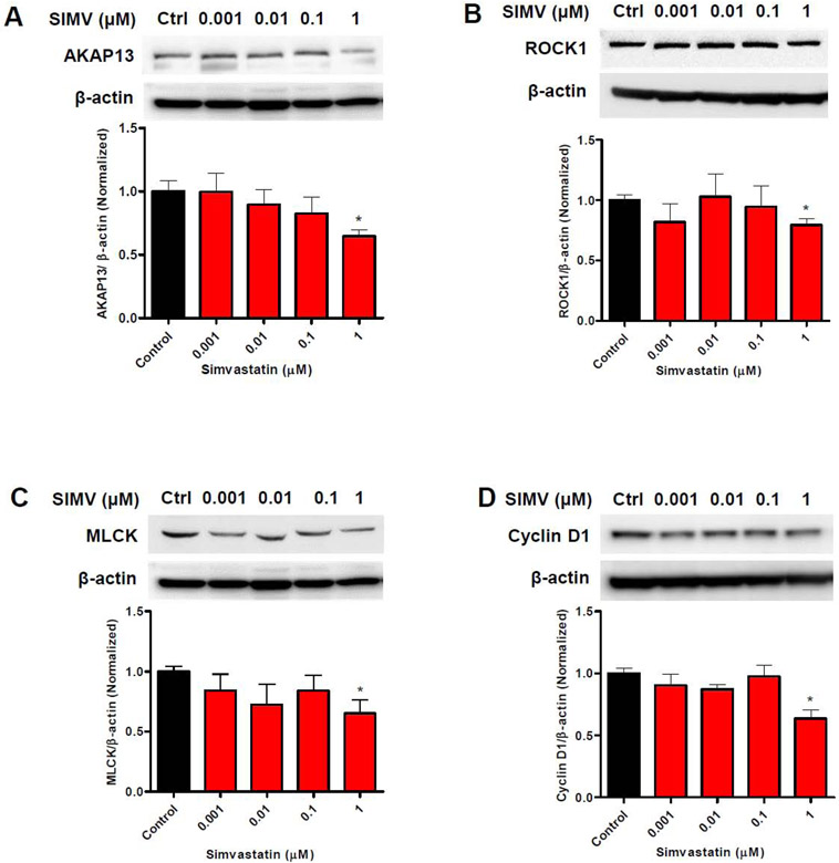
Results: We found that simvastatin significantly reduced the protein expression of β1 integrin by 44% and type I collagen by 60% compared with untreated leiomyoma cells. Simvastatin-treated cells reduced phosphorylation of focal adhesion kinase down to 26%-60% of control, whereas it increased total focal adhesion kinase protein expression. Using a Ras homolog family member A pull-down activation assay, we observed reduced levels of active Ras homolog family member A in simvastatin-treated cells by 45%-85% compared with control. Consistent with impaired Ras homolog family member A activation, simvastatin treatment reduced tumor gel contraction where gel area was 122%-153% larger than control. Furthermore, simvastatin treatment led to reduced levels of mechanical signaling proteins involved in β1 integrin downstream signaling, such as A-kinase anchor protein 13, Rho-associated protein kinase 1, myosin light-chain kinase, and cyclin D1.

Conclusion: The results of this study suggest a possible therapeutic role of simvastatin in restoring the altered state of mechanotransduction signaling in leiomyoma. Collectively, these findings are aligned with previous epidemiologic studies and other reports and support the need for clinical trials.

Keywords: AKAP13; FAK; MLCK; ROCK1; activated RhoA; cyclin D1; extracellular matrix; leiomyoma; mechanotransduction; simvastatin; type I collagen; β1 integrin.


Conflict of interest statement

Conflict of interest

William H. Catherino declares the following conflict of interest: Abbvie (Consultant); Allergan, now owned by Abbvie (Consultant); American Board of Obstetrics and Gynecology (Boards Examiner, Subject Matter Expert); American Society for Reproductive Medicine (Subject Matter Expert); Bayer (Consultant); EMD Serono (Consultant, wife is Senior Medical Director); Myovant (Consultant). Mostafa A. Borahay serves as Advisory Board member for Myovant Sciences.

Other authors report no conflict of interest

Figures

Figure 1.
Figure 1.. Altered mechanotransduction pathways in uterine leiomyoma cells.
Human leiomyoma cells surrounded by ECM consisting mainly of collagen I. The figure shows the mechanical interaction between cells and collagen I by heterodimeric (α and β1) integrin receptors. Activated β1 integrin provides mechanical forces for polymerization and organization of actin filaments and biochemical signaling for the formation of the adhesion complex triggers autophosphorylation of FAK. Phosphorylated FAK leads to the activation of AKAP13 and RhoA which further recruits ROCK1 and MLCK activation for altering mechanotransduction pathways in human leiomyoma cells. ECM, extracellular matrix; FAK, focal adhesion kinase; AKAP13, A kinase anchor protein 13; RhoA, Ras homolog gene family, member A; ROCK, RhoA kinase; MLCK, myosin light chain kinase.
Figure 2.
Figure 2.. Reduction in steady state levels of β1 integrin in simvastatin-treated leiomyoma cells.
Primary and immortalized (HuLM) human leiomyoma cells were treated with different simvastatin doses (0.001, 0.01, 0.1 and 1μM) and DMSO (control) for 48 h. The mRNA levels of β1 integrin genes were measured by quantitative real time PCR (qRT-PCR) (A, B). RPLP0 was amplified under the same qRT-PCR conditions for normalizing quantitative data. C, D: β1 integrin protein expression were determined by western blotting along with quantification and the data were normalized by β-actin. Results are expressed as mean ± standard error of the mean (SEM). *, p < 0.05; **, p < 0.01; ***, p < 0.001 versus control. The experiments were repeated three times.
Figure 3.
Figure 3.. Cytoimmunofluorescence images of β1 integrin in control and simvastatin-treated leiomyoma cells.
Primary and immortalized (HuLM) human leiomyoma cells were treated with different simvastatin doses (0.001, 0.01, 0.1 and 1μM) and DMSO (control) for 48 h. Cellular distribution of β1 integrin (green fluorescence) (A, B) were examined by immunofluorescence and confocal laser microscopy (20X imaging). Phalloidin (red fluorescence) and DAPI (blue fluorescence) were used as F-actin and nuclear markers. Scale bar = 50 μm. Fluorescence-integrated density was quantified using NIH Image J software and is presented as fold change normalized to control. *, p < 0.05; **, p < 0.01 versus control. The experiments were repeated three times.
Figure 4.
Figure 4.. Simvastatin treatment reduced phosphorylated FAK expression levels in leiomyoma cells.
Primary leiomyoma and immortalized (HuLM) human leiomyoma cells were treated with different simvastatin doses (0.001, 0.01, 0.1 and 1μM) and DMSO (control) for 48 h. The mRNA levels of FAK genes were measured by qRT-PCR (A, B). RPLP0 was amplified under the same qRT-PCR conditions for normalizing quantitative data. The protein expression level of phosphorylated to total FAK ratio (pFAK/FAK) were determined by western blotting and normalized to β-actin (C, D). Results are expressed as the mean ± SEM. *, p < 0.05; **, p < 0.01 versus control. The experiments were repeated triplicate.
Figure 5.
Figure 5.. Cytoimmunofluorescence images of FAK phosphorylation in simvastatin-treated leiomyoma cells.
Primary and immortalized (HuLM) human leiomyoma cells were treated with different simvastatin doses (0.001, 0.01, 0.1 and 1μM) and DMSO (control) for 48 h. Cellular distribution of pFAK (green fluorescence) (A, B) were examined by immunofluorescence and confocal laser microscopy (20X imaging). Phalloidin (red fluorescence) and DAPI (blue fluorescence) were used as F-actin and nuclear counterstain for all the figures. Scale bar = 50 μm. Fluorescence integrated density was quantified using NIH Image J software and is presented as fold change relative to control levels expressed as 1.00. *, p < 0.05; **, p < 0.01 versus control. The experiments were repeated three times.
Figure 6.
Figure 6.. Simvastatin treated cells showed attenuated levels of active RhoA compared to controls.
Immortalized human leiomyoma (HuLM) cells were cultured and treated with simvastatin (0.001, 0.01, 0.1, 1 and 10μM) and DMSO (control) for 48 h. Cellular lysate was made by ice-cold lysis buffer and incubated with a GST Rhotekin Rho-binding domain (GST-Rhotekin-RBD) and the activated RhoA were pulled down by beads. Western blot analysis was performed to determine the activated RhoA (GTP-bound RhoA) and the data quantified as the ratio of active to total RhoA. The 1st lane shows a 50 ng recombinant His-tagged RhoA standard (His-Rho). The subsequent 2nd to 7th lanes shows the pull-down of activated RhoA after simvastatin treatment. The 8th lane shows active GTP-loaded RhoA (GTP) and the 9th lane shows inactive GDP-loaded RhoA (GDP) from equivalent amounts of cell lysates. Results are expressed as the mean ± SEM. *, p < 0.05; **, p < 0.01 versus control. The experiments were repeated three times.
Figure 7.
Figure 7.. Reduction of gel contraction in HuLM cells after simvastatin treatment.
Immortalized human leiomyoma (HuLM) cells were treated with different simvastatin doses (0.001, 0.01, 0.1 and 1μM) and DMSO (control) for 48 h. HuLM cells were cultured in three-dimensional (3D) in collagen I gel and allowed to grow for 2 weeks before exposure of simvastatin for 96 h. Photograph were taken at the end of the treatment and the gel area was quantified by Image J software. Results are expressed as the mean ± SEM. *, p < 0.05; **, p < 0.01; ***, p < 0.001 versus control. The experiments were repeated three times.
Figure 8.
Figure 8.. Reduction of steady state mRNA transcripts encoding proteins involved in mechanical signaling following simvastatin treatment.
Immortalized human leiomyoma (HuLM) cells were treated with different simvastatin doses (0.001, 0.01, 0.1 and 1μM) and DMSO (control) for 48 h. The mRNA levels of A) AKAP13, B) ROCK1, C) MLCK and D) Cyclin D1 genes were measured by qRT-PCR. RPLP0 was amplified under the same qRT-PCR conditions for normalizing quantitative data. Results are expressed as the mean ± SEM. *, p < 0.05; **, p < 0.01; ***, p < 0.001 versus control. The experiments were repeated three times.
Figure 9.
Figure 9.. Reduction in levels of protein for key proteins involved in mechanical signaling after simvastatin treatment.
Immortalized human leiomyoma (HuLM) cells were treated with different simvastatin doses (0.001, 0.01, 0.1 and 1μM) and DMSO (control) for 48 h. The protein levels of A) AKAP13, B) ROCK1, C) MLCK and D) Cyclin D1 genes were measured by western blotting and quantified using β-actin as the loading control. Results are expressed as the mean ± SEM. *, p < 0.05; **, p < 0.01; ***, p < 0.001 versus control. The experiments were repeated three times.
Figure 10.
Figure 10.. Inhibition of type I collagen expression after simvastatin treatment.
Immortalized human leiomyoma (HuLM) cells were treated with different simvastatin doses (0.001, 0.01, 0.1 and 1μM) and DMSO (control) for 48 h. A) The mRNA level of type 1 collagen were measured by qRT-PCR. RPLP0 was amplified under the same qRT-PCR conditions for normalizing quantitative data. B) Protein expression of type 1 collagen was determined by western blotting. Data was quantified and normalized by β-actin (loading control). Results were expressed as the mean ± SEM. *, p < 0.05; **, p < 0.01; ***, p < 0.001 versus control. The experiments were repeated three times.
Figure 11.
Figure 11.. Effect of simvastatin on the mechanotransduction pathways in uterine leiomyoma cells.
A cartoon showing the beneficial effects of simvastatin on the mechanotransduction pathway. Red minus signs denote sites of simvastatin inhibitory effects.

References

    1. Humphrey JD, Dufresne ER, Schwartz MA. Mechanotransduction and extracellular matrix homeostasis. Nat Rev Mol Cell Biol 2014;15:802. - PMC - PubMed
    1. Borahay MA Al-Hendy A, Kilic GS, Boehning D. Signaling pathways in leiomyoma: understanding pathobiology and implications for therapy. Mol Med 2015;21:242. - PMC - PubMed
    1. Islam MS, Protic O, Stortoni P, et al. Complex networks of multiple factors in the pathogenesis of uterine leiomyoma. Fertil Steril 2013;100:178–93. - PubMed
    1. Bulun SE. Uterine fibroids. The New England journal of medicine 2013;369:1344–55. - PubMed
    1. Islam MS, Ciavattini A, Petraglia F, Castellucci M, Ciarmela P. Extracellular matrix in uterine leiomyoma pathogenesis: a potential target for future therapeutics. Hum Reprod Update 2018;24:59–85. - PubMed

Publication types

MeSH terms