

Longitudinal flortaucipir ([18F]AV-1451) PET uptake in semantic dementia
- PMID: 32417749
- PMCID: PMC7365267
- DOI: 10.1016/j.neurobiolaging.2020.04.010
Longitudinal flortaucipir ([18F]AV-1451) PET uptake in semantic dementia
Abstract
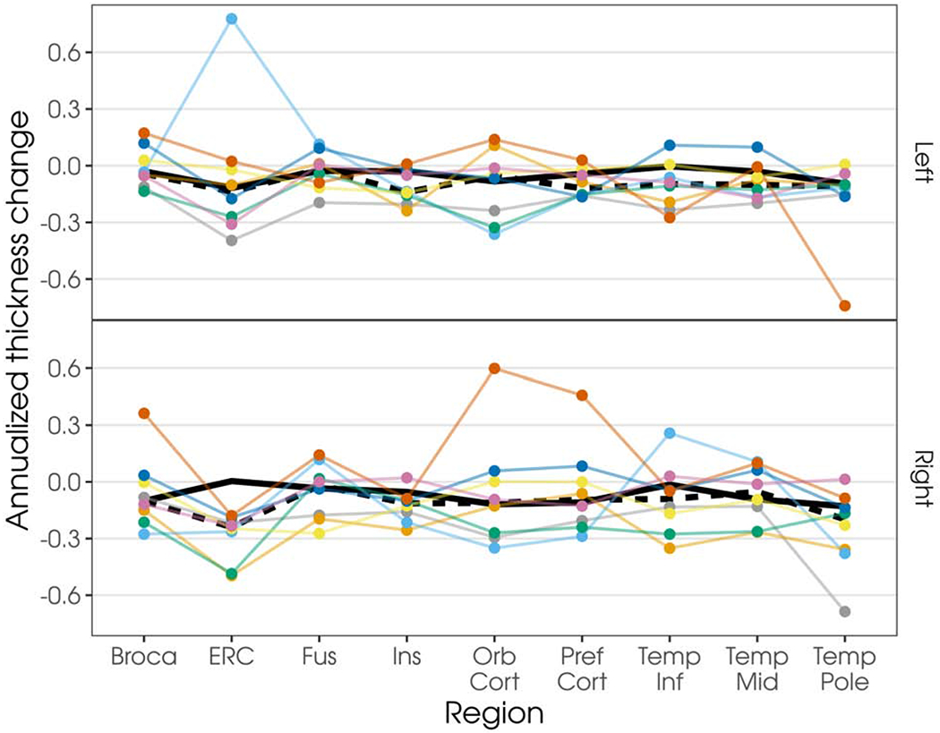
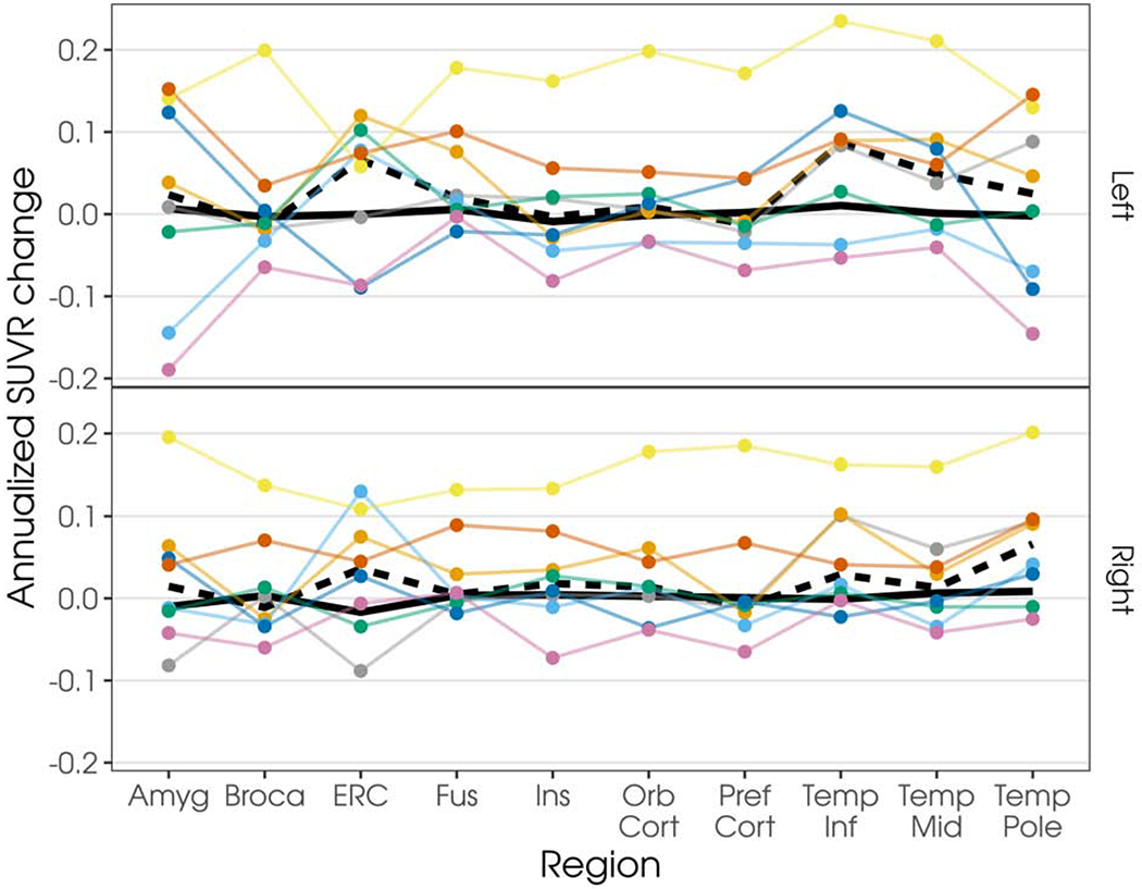
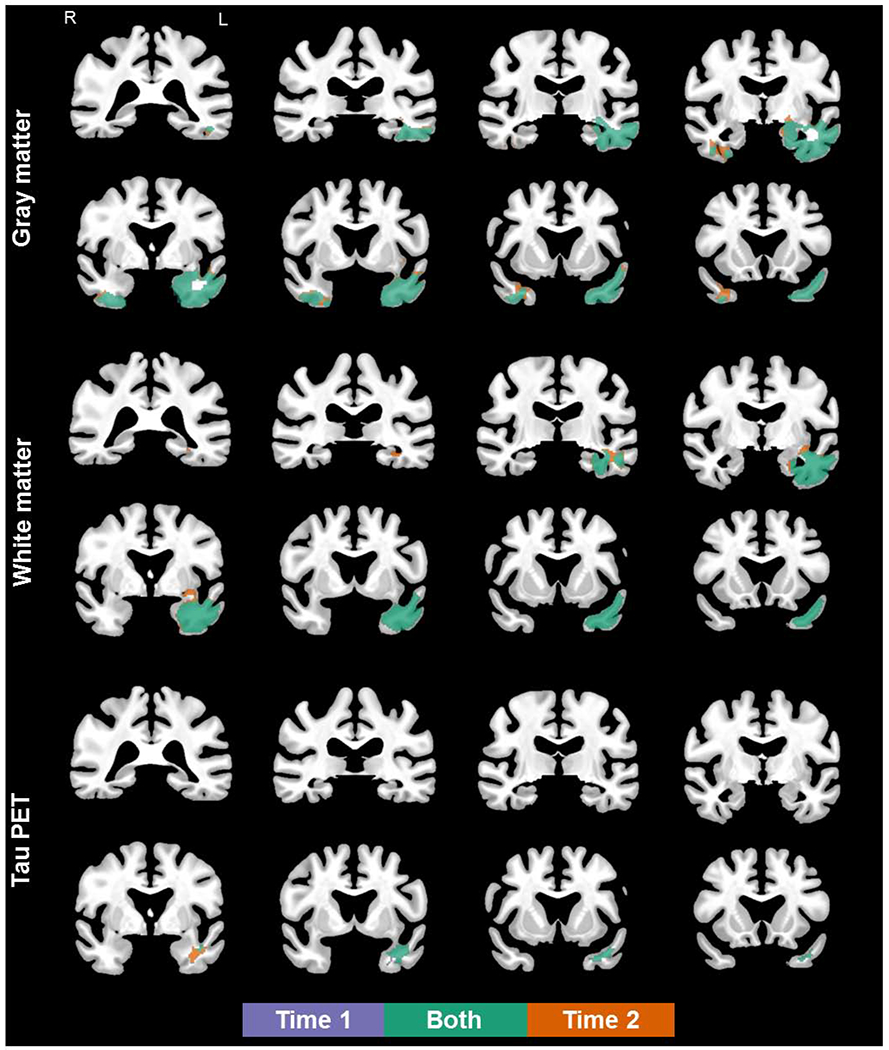
To assess volume loss and flortaucipir uptake in patients with semantic dementia (SD) over time. Eight SD patients (3 female) underwent clinical evaluations, flortaucipir positron emission tomography, and brain magnetic resonance imaging at 2 visits. Voxel-level comparisons of magnetic resonance imaging gray and white matter volume loss and flortaucipir positron emission tomography uptake were performed in SPM12, comparing SD patients to controls at each visit. T-tests on difference images and paired t-tests of flortaucipir uptake were also performed. At the voxel level, SD patients showed asymmetric, bilateral gray volume loss in the temporal lobes, which, via visual inspection, extended posteriorly at follow-up. White matter loss and flortaucipir uptake were noted in SD patients in the left temporal lobe only, which appeared to extend posteriorly, without involvement of the right hemisphere at follow-up. Longitudinal analyses did not support significant changes in flortaucipir uptake between visits. The biological mechanisms of flortaucipir signal in suspected underlying TAR-DNA binding protein 43 pathology are unknown. A 1-year interval is not sufficient time to demonstrate significant longitudinal flortaucipir uptake changes in SD.
Keywords: MRI; PET; Primary progressive aphasia; Semantic dementia; Tau.
Copyright © 2020 Elsevier Inc. All rights reserved.
Figures
References
-
- Ashburner J, & Friston KJ (2000). Voxel-Based Morphometry—The Methods. Neuroimage, 11(6), 805–821. - PubMed
-
- Ashburner J, & Friston KJ (2005). Unified segmentation. Neuroimage, 26(3), 839–851. - PubMed
-
- Bevan-Jones WR, Cope TE, Jones PS, Passamonti L, Hong YT, Fryer TD, … Rowe JB (2018). [(18)F]AV-1451 binding in vivo mirrors the expected distribution of TDP-43 pathology in the semantic variant of primary progressive aphasia. J Neurol Neurosurg Psychiatry, 89(10), 1032–1037. doi:10.1136/jnnp-2017-316402 - DOI - PMC - PubMed
Publication types
MeSH terms
Substances
Grants and funding
LinkOut - more resources
Full Text Sources
Medical
