. 2020 Dec;108(12):2504-2518.
doi: 10.1002/jbm.a.37001. Epub 2020 Jun 20.

Self-assembly of multiscale anisotropic hydrogels through interfacial polyionic complexation

Affiliations

Self-assembly of multiscale anisotropic hydrogels through interfacial polyionic complexation

Akhil Patel et al. J Biomed Mater Res A. 2020 Dec.

Abstract

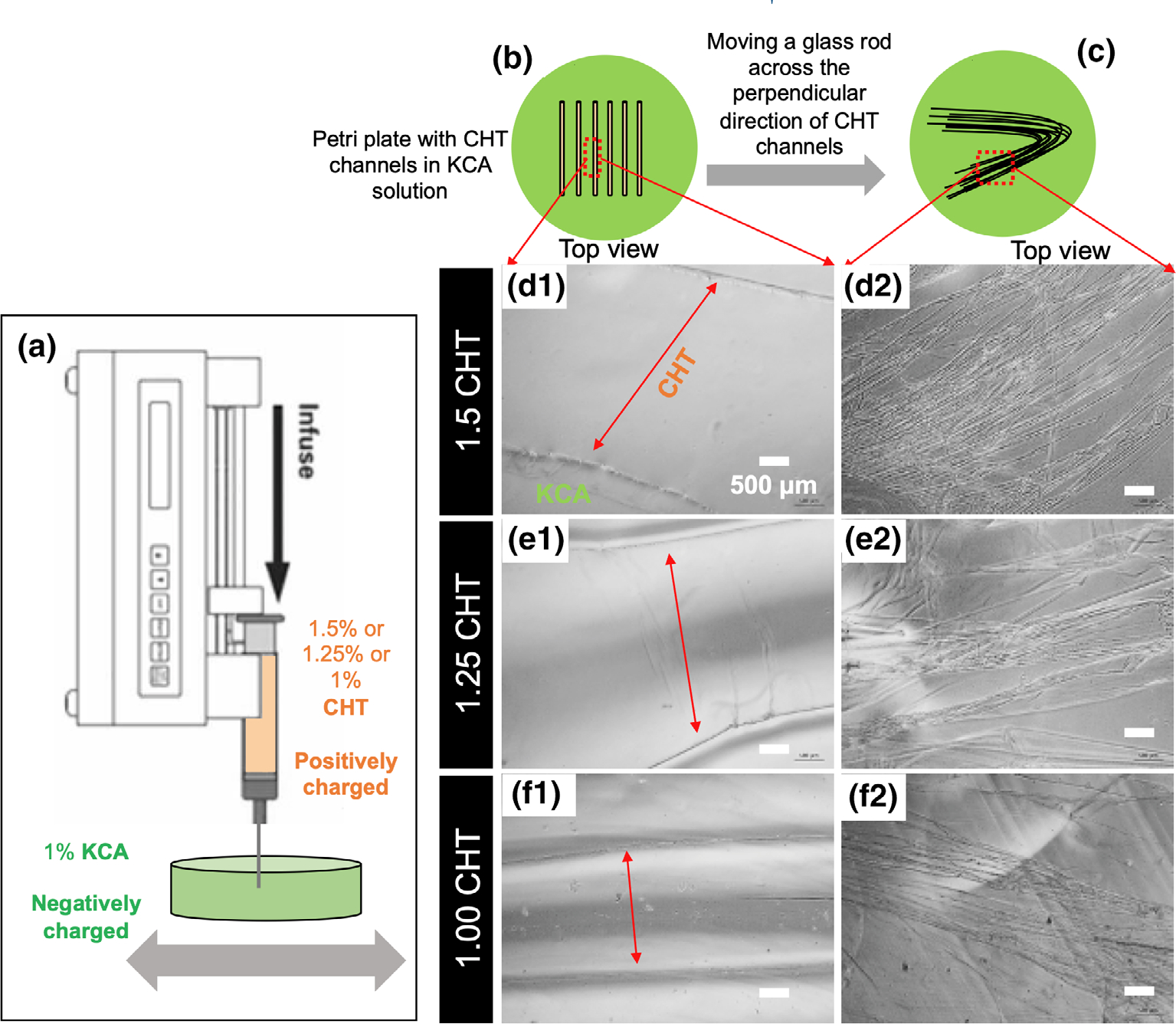
Polysaccharides are explored for various tissue engineering applications due to their inherent cytocompatibility and ability to form bulk hydrogels. However, bulk hydrogels offer poor control over their microarchitecture and multiscale hierarchy, parameters important to recreate extracellular matrix-mimetic microenvironment. Here, we developed a versatile platform technology to self-assemble oppositely charged polysaccharides into multiscale fibrous hydrogels with controlled anisotropic microarchitecture. We employed polyionic complexation through microfluidic flow of positively charged polysaccharide, chitosan, along with one of the three negatively charged polysaccharides: alginate, gellan gum, and kappa carrageenan. These hydrogels were composed of microscale fibers, which in turn were made of submicron fibrils confirming multiscale hierarchy. Fibrous hydrogels showed strong tensile mechanical properties, which were further modulated by encapsulation of shape-specific antioxidant cerium oxide nanoparticles (CNPs). Specifically, hydrogels with chitosan and gellan gum showed more than eight times higher tensile strength compared to the other two pairs. Incorporation of sphere-shaped cerium oxide nanoparticles in chitosan and gellan gum further reinforced fibrous hydrogels and increased their tensile strength by 40%. Altogether, our automated hydrogel fabrication platform allows fabrication of bioinspired biomaterials with scope for one-step encapsulation of small molecules and nanoparticles without chemical modification or use of chemical crosslinkers.

Keywords: alginate; automated collector; cerium oxide nanoparticles; chitosan; fibrous hydrogels; gellan gum; interfacial polyionic complexation; kappa carrageenan; polysaccharides.


Conflict of interest statement

CONFLICT OF INTEREST

The authors declare no potential conflict of interest.

Figures

FIGURE 1
FIGURE 1
Physical characterization and bulk mixing of oppositely charged polysaccharide solutions. Table 1 shows the zeta potential values of aqueous solutions of polysaccharides; (1a) oppositely charged polysaccharide pairs with arrows; (a1–a4) effect of varying concentrations (% wt/vol) of chitosan (CHT) (a1) alginic acid (a2), gellan gum (GG) (a3), and kappa carrageenan (KCA) (a4) on the viscosity at various shear rates (n = 3 per concentration). Concentration showed significant difference in two-way analysis of variance analysis for all four polysaccharides. Shear rates showed significant difference only for CHT and KCA solutions, y-axis shows logarithmic scale; (b1, b2) representative light microscopic images of crosslinked fibrous and nonfibrous complexes resulted after bulk mixing of oppositely charged polysaccharide solutions. (c) Bulk mixing of oppositely charged polysaccharide solutions on a XY plate shaker leading to encapsulation of one of the solutions inside the electrostatically crosslinked membranous interface
FIGURE 2
FIGURE 2
Interfacial mixing of oppositely charged polysaccharide solutions. Droplets of chitosan (CHT) and kappa carrageenan (KCA) (a1) were brought in contact with each other to form electrostatically crosslinked interface (a2). Pipet was used to mix gently at the interface (a3) and small amount was pulled away at the interface (a4) resulting in an interfacial fiber (a5). (a6) Schematic representation of interfacial mixing of oppositely charged polysaccharide solutions resulting in formation of fibrous complexes. (b1–b3) Scanning electron microscopy images of single fiber of CHT-alginic acid (ALG), CHT-gellan gum (GG), and CHT-KCA, respectively; (b4) fibril diameter quantification for the three types of hydrogels, n ≥ 10 fibers per image, three images per group, * indicates p < 0.05 (one-way analysis of variance and Tukey’s post-hoc test). (c) XY matrix showing effect of polymer concentration on the form of complexes that dominate (fibrous or nonfibrous) after interfacial mixing for all three pairs, CHT + ALG, CHT + GG, and CHT + KCA. (d1, d2; e1, e2; and f1, f2) Light microscopy images of complexes as a result of interfacial mixing of CHT + ALG, CHT + GG, and CHT + KCA, respectively. 1% wt/vol combinations showed fibrous complexes whereas 0.5% wt/vol combinations showed dominant nonfibrous complexes in light microscopy images. (d3, e3, and f3) Absorbance values at 630 nm for turbid complexes as a result of interfacial mixing of various concentration of CHT + ALG, CHT + GG, and CHT + KCA, respectively. Combination of all three pairs at 1% wt/vol of each polysaccharide and that of KCA (1% wt/vol) with CHT (1.25 or 1.5% wt/vol) showed relatively higher absorbance values as compared to other concentration and solution pairs
FIGURE 3
FIGURE 3
Three-dimensional printing of chitosan (CHT) channels in kappa carrageenan (KCA) solution. (a) Set up with inverted syringe pump programmed at 50 ml/hr dispensing either CHT (1, 1.25, or 1.5% wt/vol) into KCA (1% wt/vol) solution at 60°C in a manually movable petri plate. (b) Top view of petri plate after printing CHT channels parallel to each other. (c) Fiber bundles formed by moving a glass rod across CHT channels in the perpendicular direction; (d1, e1, f1) as-printed CHT channels (1.5, 1.25, and 1% wt/vol) in KCA (1% wt/vol). (d2, e2, f2) Formation of fiber bundles as transparent fibers with glossy boundaries
FIGURE 4
FIGURE 4
Interfacial mixing chamber with automated fiber collector assembly. (a1) A schematic of the set up for automated collection of fibers in a temperature-controlled incubator using two types of motors: one moving in the circular direction to collect and cut fibers on a glass slide while the other (programmable stage) moving along the horizontal axis to arrange fibers parallel to each other; red-dotted outline pictures exhibit corresponding area in the schematic (red dotted outline) before (a2) and after (a3) collection of the fiber. (b1) Assembly with both the motors and fiber dispensing needle in the controlled temperature incubator; (b2) coverslip near fiber dispensing needle; (c1) effect of pump speed on formation of fibers per minute and fiber formation per milliliter; (c2) effect of stage speed on fiber distribution density (fibers/cm2) and hydrogel scaffold area covered per unit volume (cm2/ml), respectively; * and # indicates p < 0.05 (one-way analysis of variance and Tukey’s post-hoc test). (d1, d2) Representative color survey corresponding to the fiber orientation angle output images from FIJI of light microscopy images from manual and automated collector hydrogels, respectively; red color indicates parallel fibers. (d3) Histogram showing distribution of angles from hydrogels made by at least two users independently using either manual (blue) and automated (green) collector; 90° refers to the parallel alignment of fibers. The inset bar graphs show SD for each user from the histograms for corresponding methods. Note higher differences in SD for manual collector (blue bar graphs in the inset)
FIGURE 5
FIGURE 5
Cytocompatibility of fibrous hydrogels. (a) Cytocompatibility of MC3T3-mouse preosteoblast cells seeded on hydrogels at days 1, 3, 7, and 14, measured by metabolic activity alamarBlue assay. There was no statistical significance between hydrogel conditions at the same time point; (b) cytoskeletal images of MC3T3 cells seeded and cultured for 14 days on chitosan-kappa carrageenan hydrogels; actin: green and nuclei: blue
FIGURE 6
FIGURE 6
Uniaxial tensile mechanical properties of all three types of hydrogels. (a–d) Mechanical properties of chitosan (CHT)-gellan gum, CHT-kappa carrageenan, and CHT-alginic acid hydrogels showing Young’s modulus (a), percentage elongation (b), ultimate tensile strength (c), and toughness comparison (d); n = 3; *p < 0.05 (one-way analysis of variance and Tukey’s post-hoc test)
FIGURE 7
FIGURE 7
Uniaxial tensile mechanical properties of cerium oxide nanoparticle (CNP)-chitosan (CHT)-gellan gum (GG) hydrogels. (a–d) Mechanical properties of CHT-GG hydrogels encapsulating sphere- or rod-shaped CNPs (5% wt/vol) (referred as CNP-CHT-GG), showing Young’s modulus (a), percentage elongation (b), ultimate tensile strength (c), and toughness comparison (d); n = 3; *p < 0.05; one-way analysis of variance and Tukey’s post-hoc test

References

    1. Abbah SA, Liu J, Lam RWM, Goh JCH, & Wong HK (2012). In vivo bioactivity of rhBMP-2 delivered with novel polyelectrolyte complexation shells assembled on an alginate microbead core template. Journal of Controlled Release, 162(2), 364–372. - PubMed
    1. Amaike M, Senoo Y, & Yamamoto H (1998). Sphere, honeycomb, regularly spaced droplet and fiber structures of polyion complexes of chitosan and gellan. Macromolecular Rapid Communications, 19(6), 287–289.
    1. Annabi N, Shin SR, Tamayol A, Miscuglio M, Bakooshli MA, Assmann A, … Khademhosseini A (2016). Highly elastic and conductive human-based protein hybrid hydrogels. Advanced Materials, 28(1), 40–49. - PMC - PubMed
    1. Arenales-Sierra IM, Lobato-Calleros C, Vernon-Carter EJ, Hernandez-Rodriguez L, & Alvarez-Ramirez J (2019). Calcium alginate beads loaded with Mg(OH)(2) improve L. casei viability under simulated gastric condition. Lwt-Food Science and Technology, 112.
    1. Bidarra SJ, Barrias CC, & Granja PL (2014). Injectable alginate hydrogels for cell delivery in tissue engineering. Acta Biomaterialia, 10, 1646–1662. - PubMed

Publication types

LinkOut - more resources