Review
. 2021 Feb:110:2-10.
doi: 10.1016/j.semcdb.2020.04.017. Epub 2020 May 14.

Role of cilia in the pathogenesis of congenital heart disease

Affiliations
Review

Role of cilia in the pathogenesis of congenital heart disease

George C Gabriel et al. Semin Cell Dev Biol. 2021 Feb.

Abstract

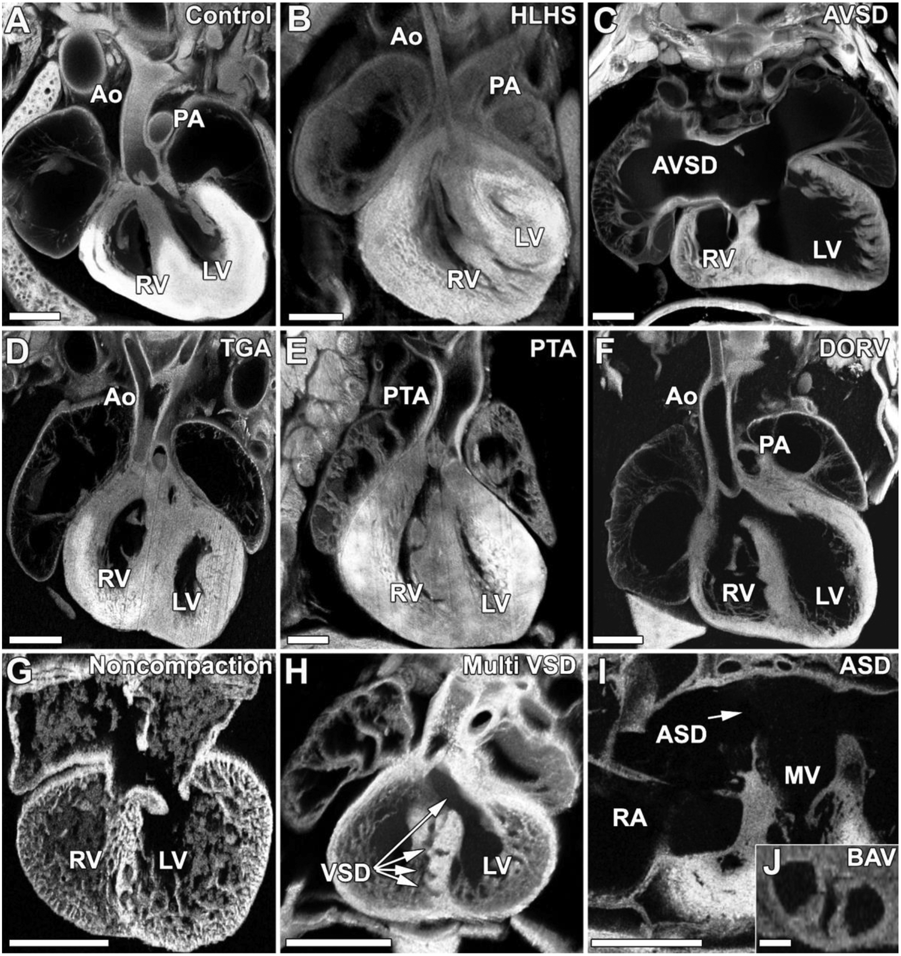
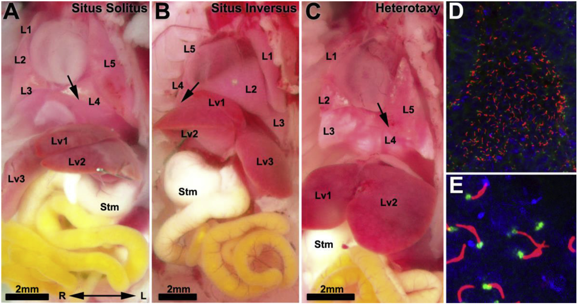
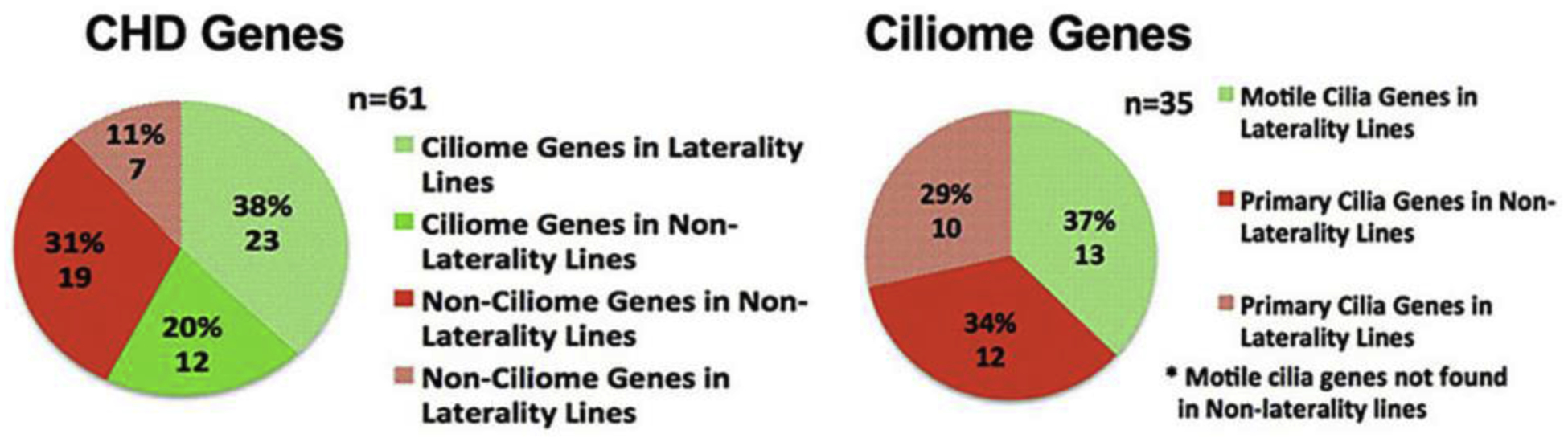
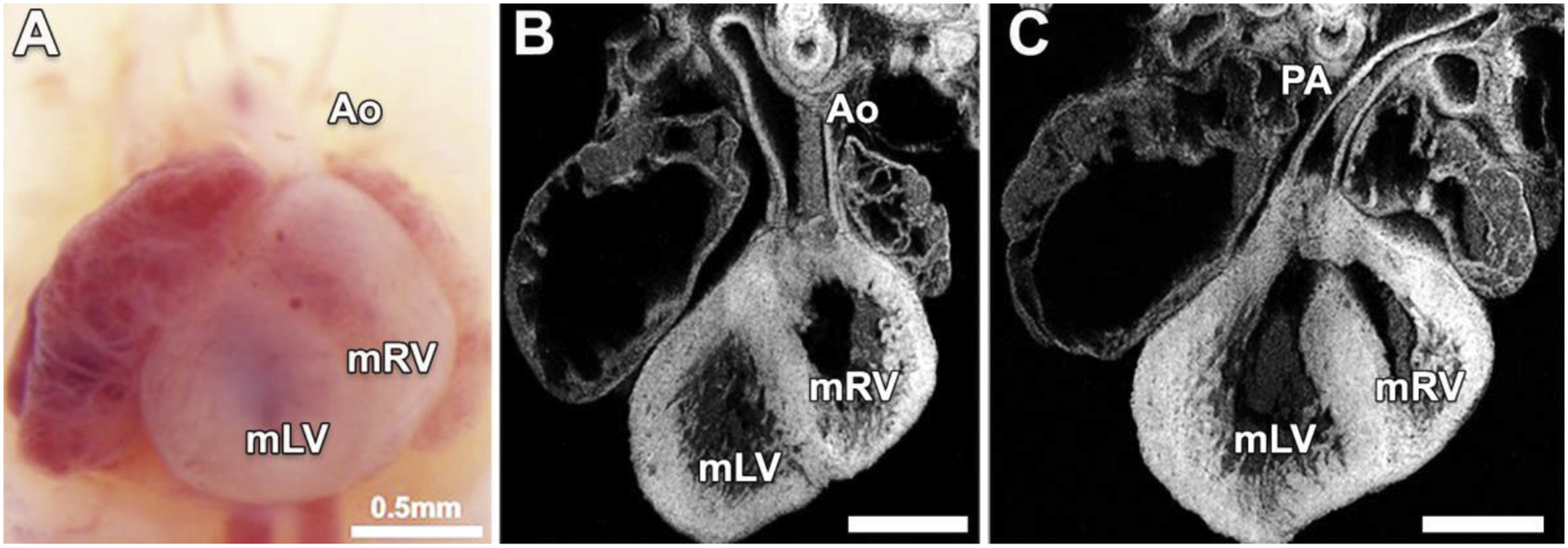
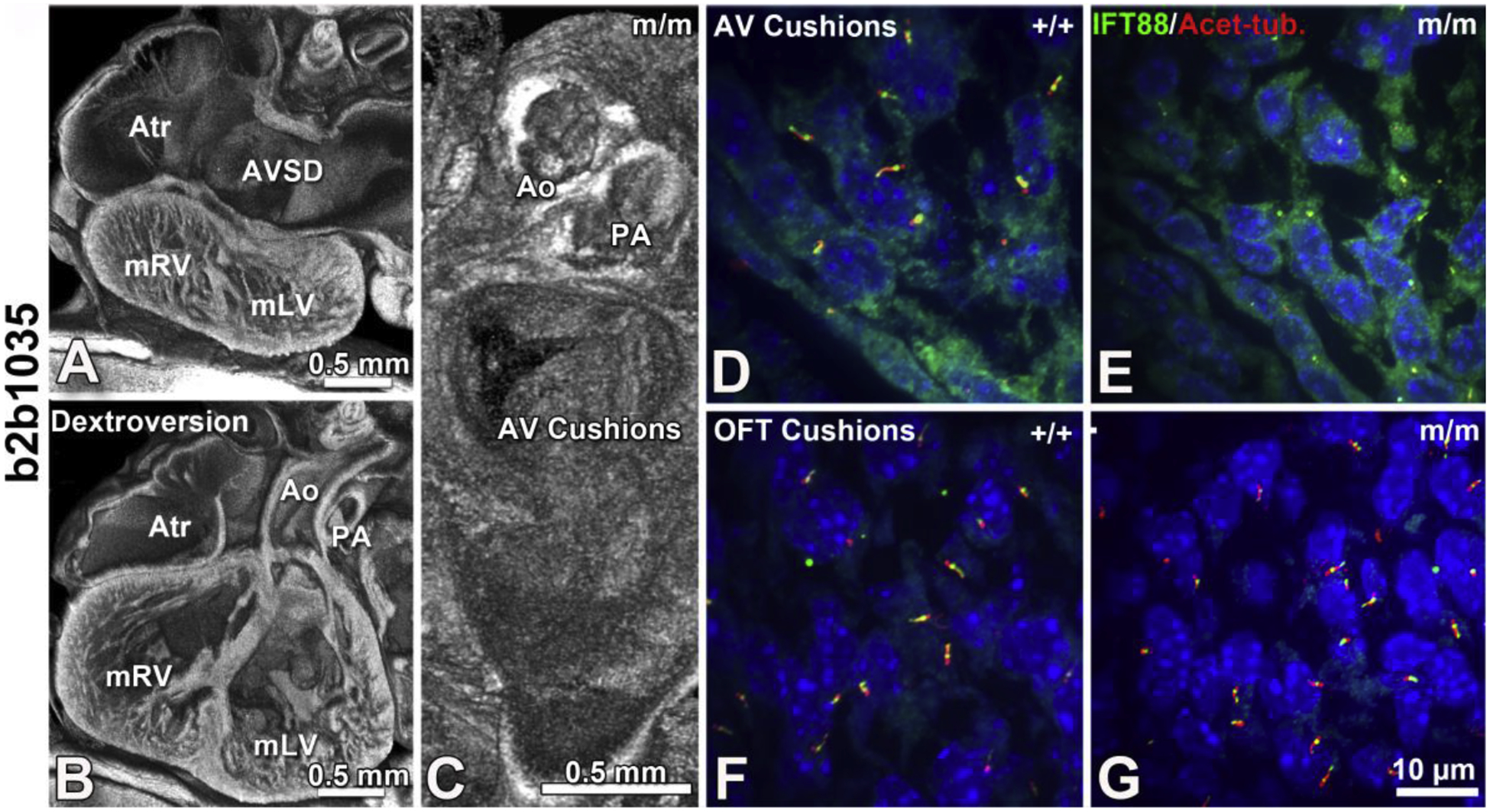
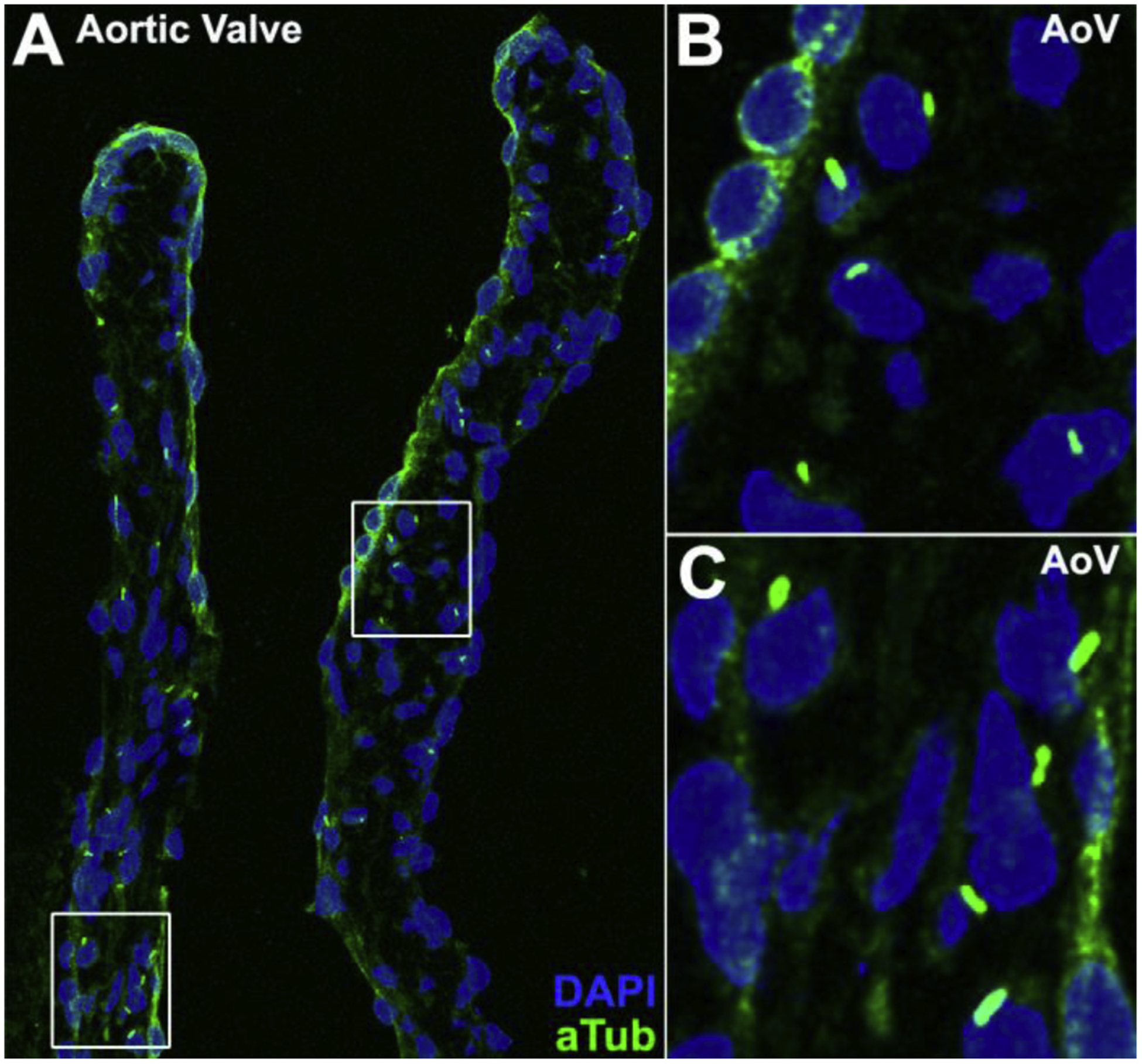
An essential role for cilia in the pathogenesis of congenital heart disease (CHD) has emerged from findings of a large-scale mouse forward genetic screen. High throughput screening with fetal ultrasound imaging followed by whole exome sequencing analysis recovered a preponderance of cilia related genes and cilia transduced cell signaling genes among mutations identified to cause CHD. The perturbation of left-right patterning in CHD pathogenesis is suggested by the association of CHD with heterotaxy, but also by the finding of the co-occurrence of laterality defects with CHD in birth defect registries. Many of the cilia and cilia cell signaling genes recovered were found to be related to Hedgehog signaling. Studies in mice showed cilia transduced hedgehog signaling coordinates left-right patterning with heart looping and differentiation of the heart tube. Cilia transduced Shh signaling also regulates later events in heart development, including outflow tract septation and formation of the atrioventricular septum. More recent work has shown mutations in cilia related genes may also contribute to valve disease that largely manifest in adult life. Overall, these and other findings show cilia play an important role in CHD and also in more common valve diseases.

Keywords: Cilia; Congenital heart disease; Fetal ultrasound imaging; Forward genetics; Mouse models; Mutagenesis.


Conflict of interest statement

Conflict of interest statement

The authors declare no conflict of interest.

Figures

Figure 1.
Figure 1.. Wide spectrum of congenital heart defect phenotypes observed in mutant mice recovered from the forward genetic screen.
Mutant mice recovered from the mutagenesis screen exhibit a wide range of congenital heart disease phenotypes. For comparison a normal mouse heart is shown in (A). In panels (B-J) are shown a wide range of CHD phenotypes recovered from the screen including hypoplastic left heart syndrome (HLHS) in which left sided heart structures are underdeveloped (B), atrioventricular septal defect in which the septa between the right and left atria and the right and left ventricles fail to form properly (C), transposition of the great arteries (TGA) in which the aorta exits the right ventricle while the pulmonary artery emerges from the left ventricle (D), persistent truncus arteriosis (PTA) in which a single outflow tract arises from the ventricles (E), double outlet right ventricle (DORV) in which both the aorta and pulmonary arteries arises from the right ventricle (F), ventricular noncompaction in which the compact myocardium fails to form appropriately (G), swiss cheese heart in which multiple ventricular septal defects (VSD) are present (H), atrial septal defect (ASD) in which the atrial septum fails to form appropriately (I), or bicuspid aortic valve (BAV) in which the normally tricuspid aortic valve develops with only two cusps (J). Ao, aorta; PA, pulmonary artery; LV, left ventricle; RV, right ventricle; MV, mitral valve.
Figure 2.
Figure 2.. Mutations causing laterality defects can result in three situs phenotypes
(A–C). Laterality mutants can present with three phenotypes: situs solitus with normal visceral organ patterning in which the heart apex points to the left with four right lung lobes and one left lung lobe, and the dominant liver lobe on the right (A); situs inversus with complete mirror reversal of visceral organ patterning (B); or heterotaxy, in which there is randomization of visceral organ patterning such that the heart apex in this mutant points leftward while the stomach (Stm) is positioned on the right (C). L1-L5, lung lobes 1–5; Lv1-Lv3, liver lobes 1–3; stm, stomach. (D,E). Motile cilia in the embryonic node are visualized with acetylated tubulin (red) labeing the ciliary axoneme and gamma tubulin (green) labeling the basal body.
Figure 3.
Figure 3.. Pathogenic mutations in ciliome genes enriched in mouse ENU mutagenesis screen
Exome sequencing from 113 CHD mutant mouse lines recovered 91 pathogenic mutations in 61 genes. 35 of these mutations affected cilia genes and caused CHD including 23 in lines exhibiting CHD with laterality defects, and 12 in lines without laterality defects. Note mutations in motile cilia genes were recovered only in mutant lines with laterality defects. Adapted from Li et al. Nature 2015 [13].
Figure 4.
Figure 4.. Megf8 mutant mice with dextrocardia and transposition of the great arteries.
Necropsy image (A) and histological sections of the same heart (B,C) from a Megf8 mutant mouse show dextrocardia with transposition of the great arteries (TGA). Note the heart apex points to the right, with the aorta (Ao) connected to the morphological right ventricle (mRV) located on the anatomical left (B), and the pulmonary artery (PA) connected to the morphological left ventricle (mLV) located on the anatomical right (C).
Figure 5.
Figure 5.. Cc2d2a mutant mice with laterality defects show AVSD with cilia defects in the AV cushion mesenchyme.
Mice with mutations in Cc2d2a present with laterality defects and congenital heart disease including dextroversion and atrioventricular septal defects (AVSD, A,B) with malformation of the atrioventricular cushions but normal outflow tract patterning (C). Consistent with this Cc2d2a mutant mice also show reduced cilia in the AV cushions compared to control (D vs E), but normal ciliation in the outflow tract cushions (F vs G). Adapted from Li et al. Nature 2015 [13].
Figure 6.
Figure 6.. Primary cilia in adult mouse aortic valve
Primary cilia are observed in the aortic valve of a 3-month-old C57BL6/J mouse with immunostaining using an acetylated tubulin antibody (A). Boxed regions in panel (A) are shown in enlarged views in panels (B) and (C).

References

    1. Liu Y, Chen S, Zuhlke L, Black GC, Choy MK, Li N, et al., Global birth prevalence of congenital heart defects 1970–2017: updated systematic review and meta-analysis of 260 studies, Int J Epidemiol (2019). - PMC - PubMed
    1. Longobardo L, Jain R, Carerj S, Zito C, Khandheria BK, Bicuspid aortic valve: unlocking the morphogenetic puzzle, The American journal of medicine 129(8) (2016) 796–805. - PubMed
    1. Tutar E, Ekici F, Atalay S, Nacar N, The prevalence of bicuspid aortic valve in newborns by echocardiographic screening, American heart journal 150(3) (2005) 513–515. - PubMed
    1. Gupta A, Amin Z, Popular Hybrid Congenital Heart Procedures without Cardiopulmonary Bypass, Frontiers in surgery 4 (2017) 9. - PMC - PubMed
    1. Holst KA, Said SM, Nelson TJ, Cannon BC, Dearani JA, Current interventional and surgical management of congenital heart disease: specific focus on valvular disease and cardiac arrhythmias, Circulation research 120(6) (2017) 1027–1044. - PubMed

Publication types

MeSH terms