. 2020 Aug;73(8):1473-1481.
doi: 10.1016/j.bjps.2020.03.012. Epub 2020 Apr 11.

Introducing human adipose-derived mesenchymal stem cells to Avance nerve grafts and NeuraGen nerve guides

Affiliations

Introducing human adipose-derived mesenchymal stem cells to Avance nerve grafts and NeuraGen nerve guides

Femke Mathot et al. J Plast Reconstr Aesthet Surg. 2020 Aug.

Abstract

Background: When direct nerve coaptation is impossible after peripheral nerve injury, autografts, processed allografts, or conduits are used to bridge the nerve gap. The purpose of this study was to examine if human adipose-derived Mesenchymal Stromal/Stem Cells (MSCs) could be introduced to commercially available nerve graft substitutes and to determine cell distribution and the seeding efficiency of a dynamic seeding strategy.

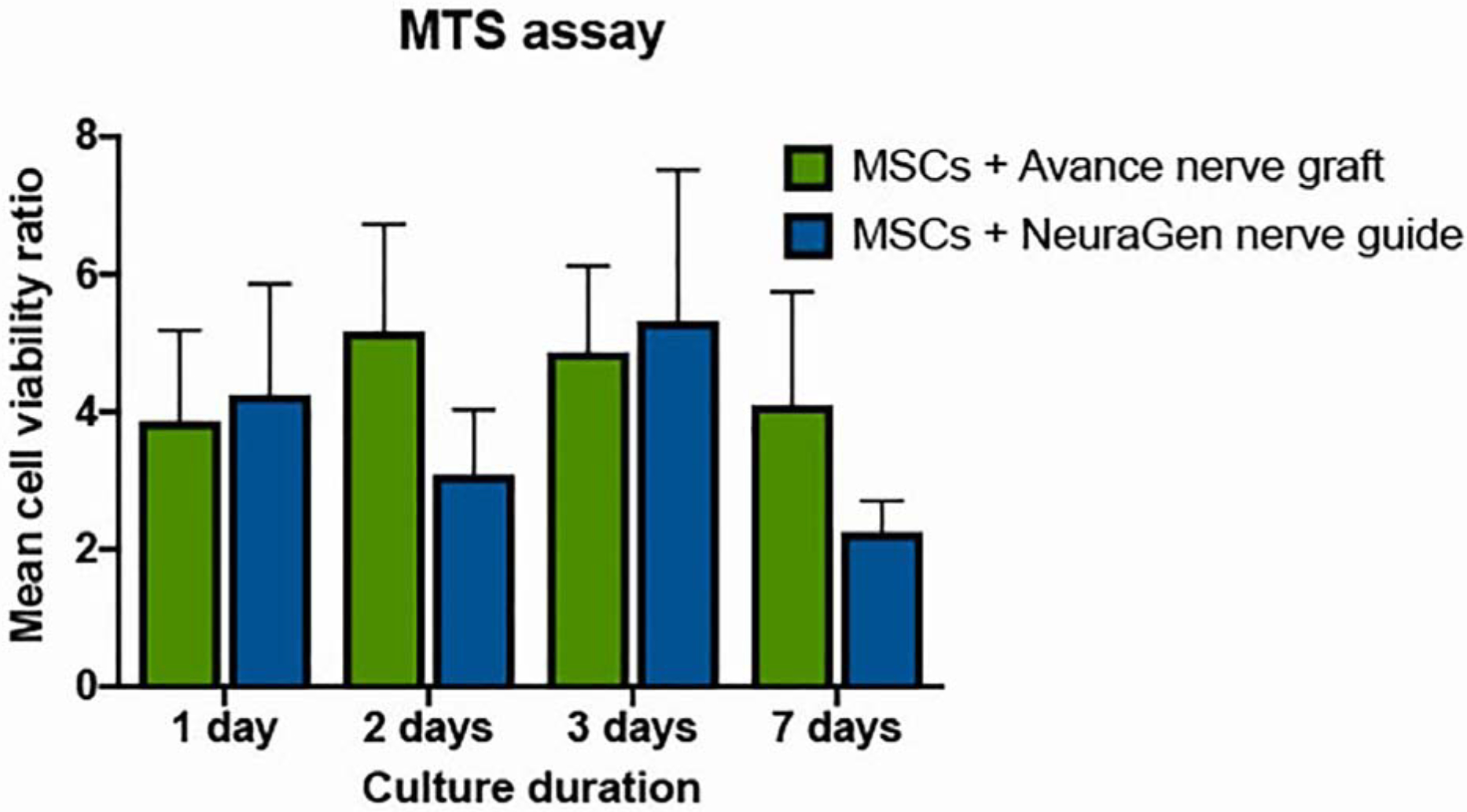
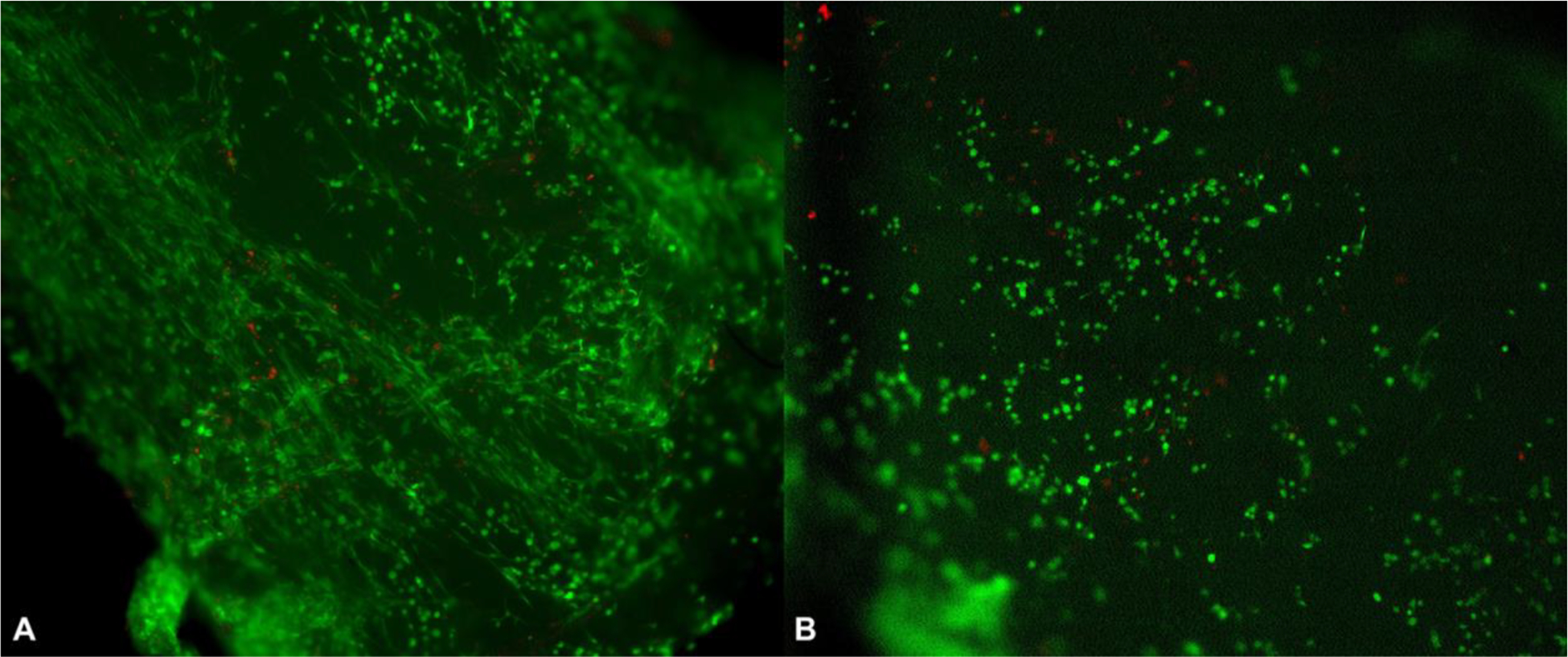
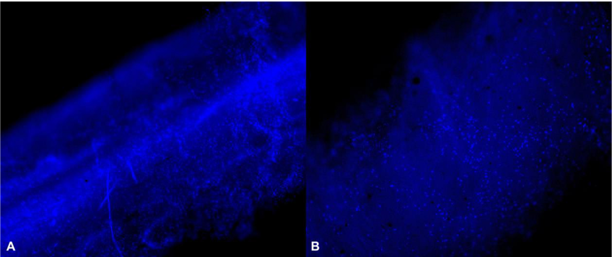
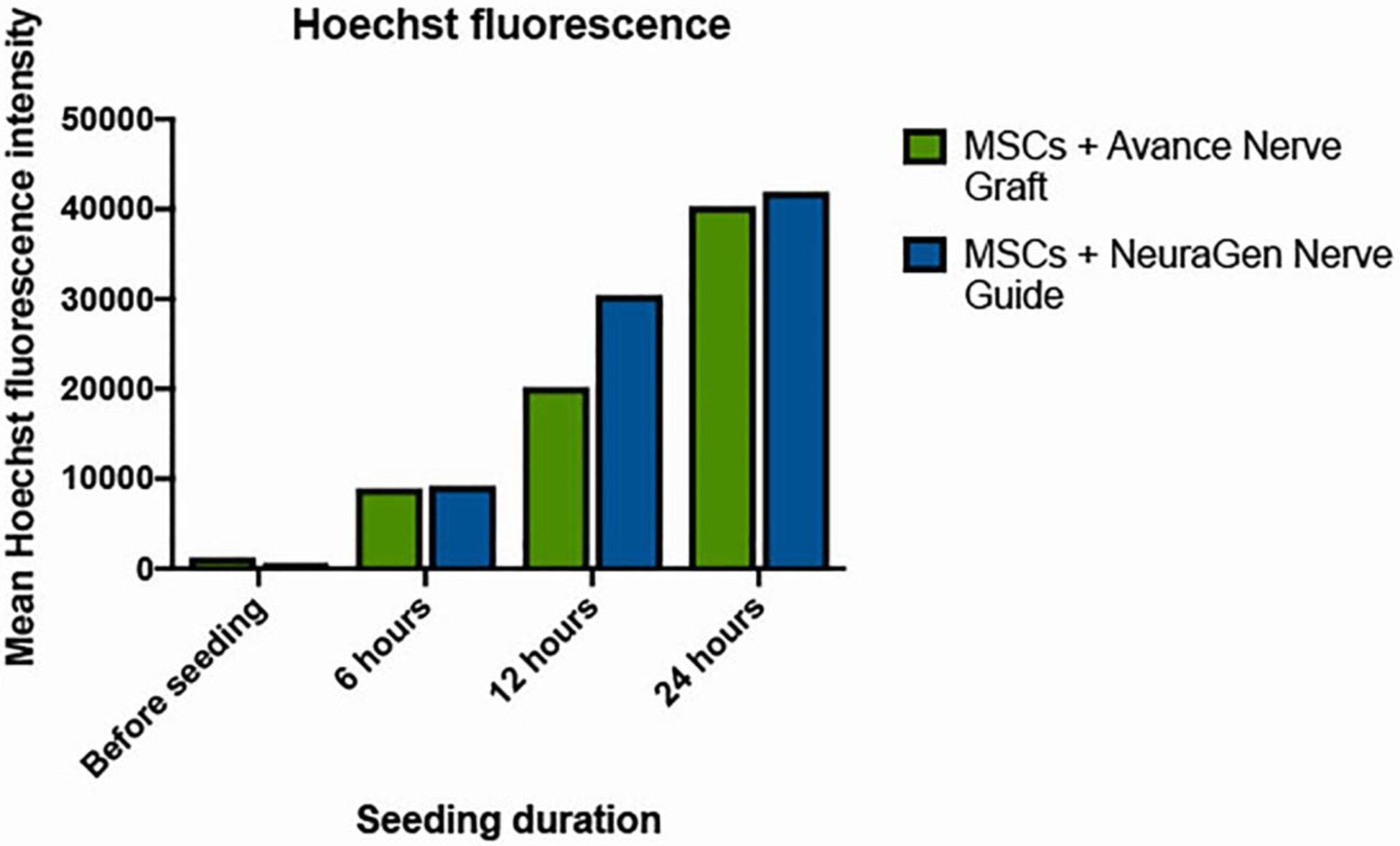
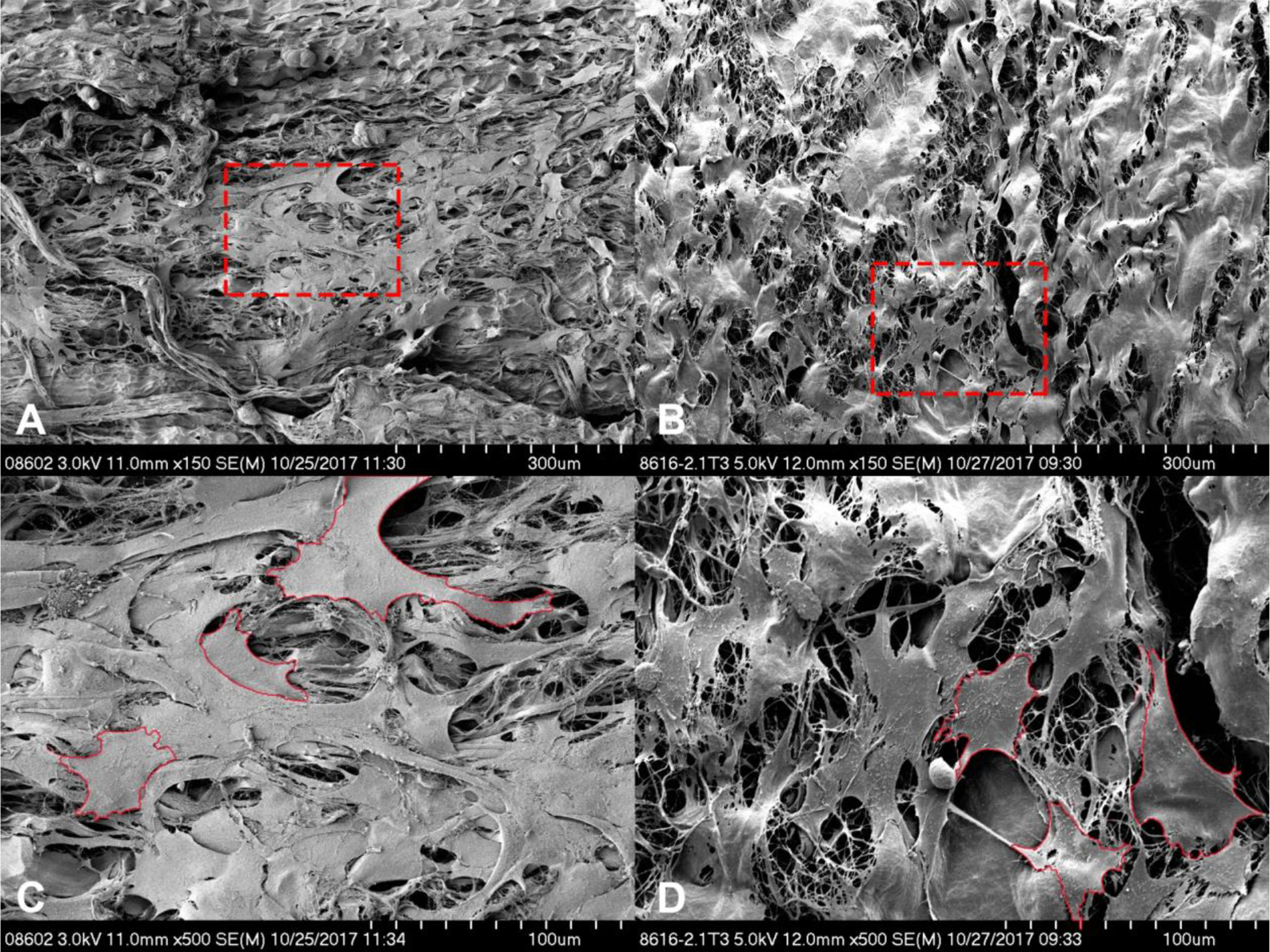
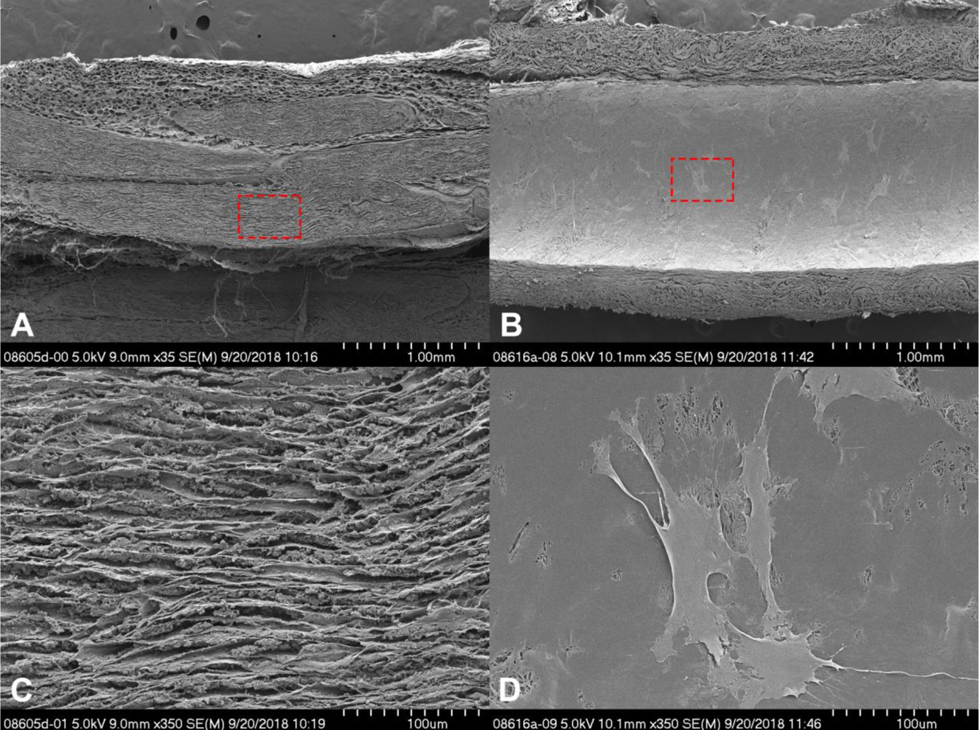
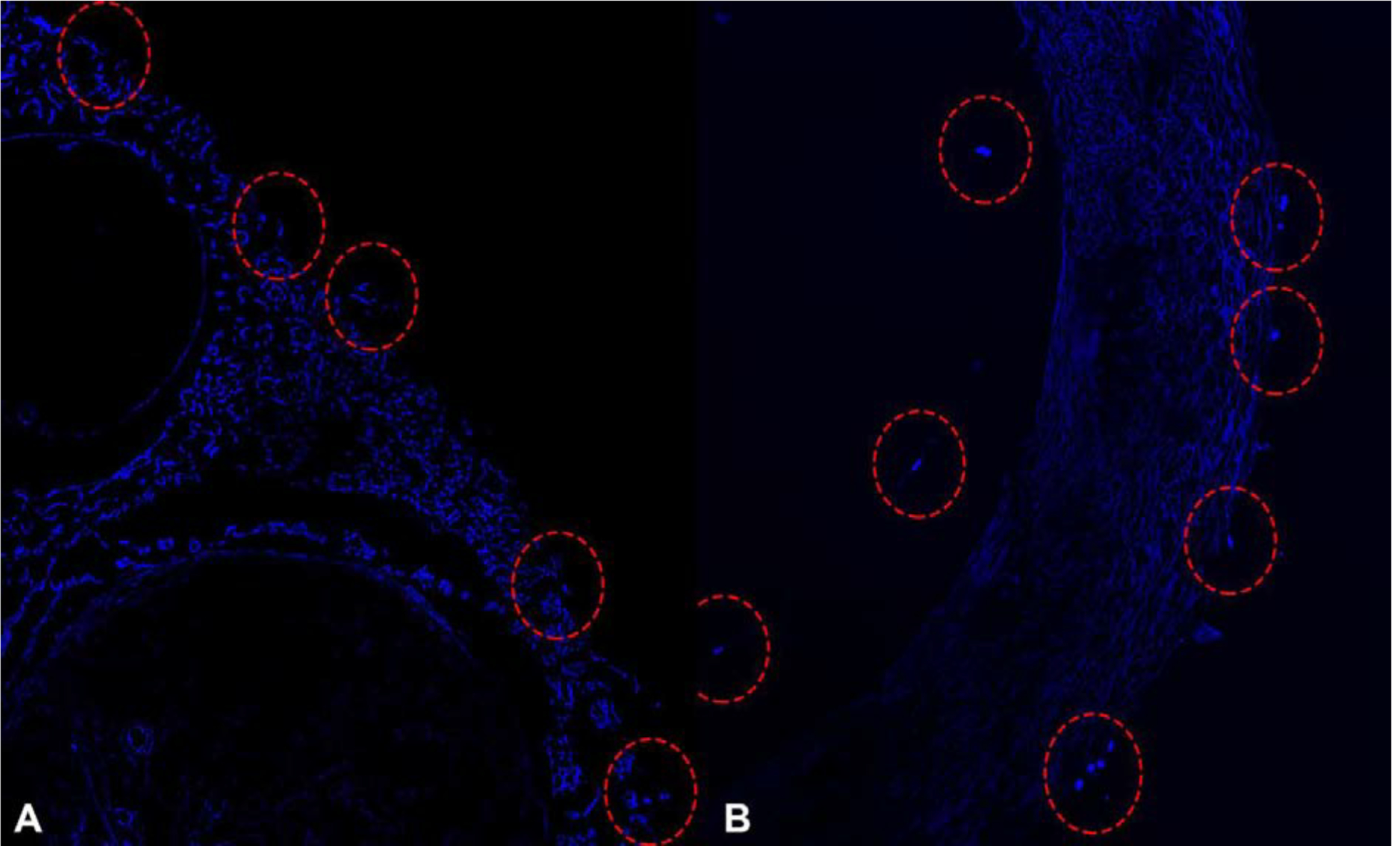
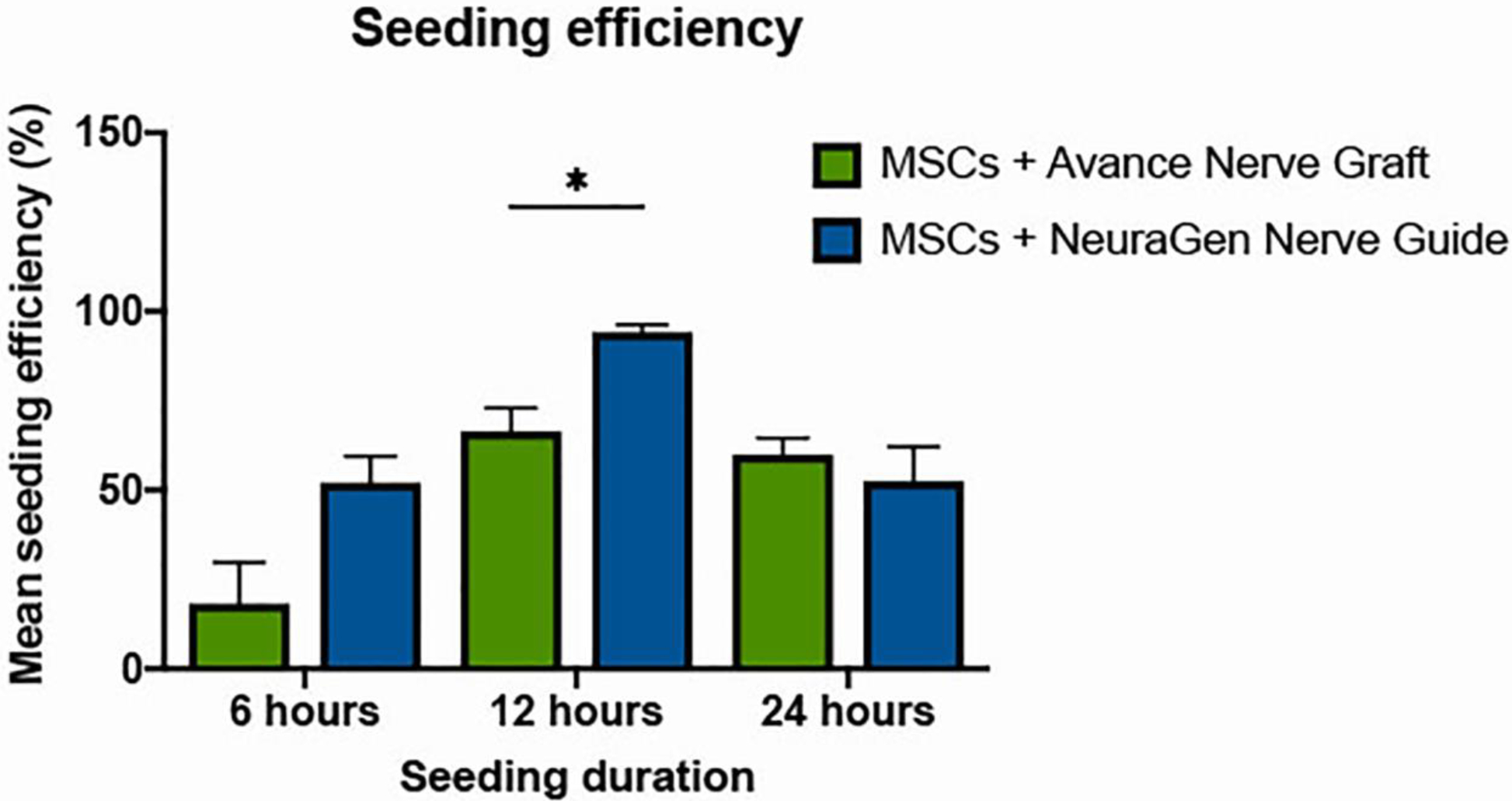
Methods: MTS assays examined the viability of human MSCs after introduction to the Avance Nerve Graft and the NeuraGen Nerve Guide. MSCs were dynamically seeded on nerve substitutes for either 6, 12, or 24 h. Cell counts, live/dead stains, Hoechst stains, and Scanning Electron Microscopy (SEM) revealed the seeding efficiency and the distribution of MSCs after seeding.

Results: The viability of MSCs was not affected by nerve substitutes. Dynamic seeding led to uniformly distributed MSCs over the surface of both nerve substitutes and revealed MSCs on the inner surface of the NeuraGen Nerve Guides. The maximal seeding efficiency of NeuraGen Nerve Guides (94%), obtained after 12 h was significantly higher than that of Avance Nerve Grafts (66%) (p = 0.010).

Conclusion: Human MSCs can be dynamically seeded on Avance Nerve Grafts and NeuraGen Nerve Guides. The optimal seeding duration was 12 h. MSCs were distributed in a uniform fashion on exposed surfaces. This study demonstrates that human MSCs can be effectively and efficiently seeded onto commercially available nerve autograft substitutes in a timely fashion and sets the stage for the clinical application of MSC-seeded nerve graft substitutes clinically.

Keywords: Avance(Ⓡ) Nerve Graft; MSCs; NeuraGen(Ⓡ) Nerve Guide; Seeding.


Conflict of interest statement

Declaration of Competing Interest The authors have nothing to disclose. This study was funded by the NIH R01, “Bridging the gap: angiogenesis and stem cell seeding of processed nerve allograft” 1 RO1 NS102360-01A1. The Avance(Ⓡ) Nerve Grafts used in this study were provided by AxoGen Inc., Alachua, Florida, USA. The NeuraGen(Ⓡ) Nerve Guides used in this study were provided by Integra LifeSciences Holdings Corporation, Plainsboro, New Jersey, USA.

Figures

Figure 1.
Figure 1.
Cell viability over time of MSCs when combined with the Avance® Nerve Graft and the NeuraGen® Nerve Guide (n=3 per group per time point). Viability of MSCs in the presence of the Avance® Nerve Graft and the NeuraGen® Nerve Guide is expressed as a ratio of the viability of MSCs without any of the nerve substitutes. pHEMA coating was used in all groups presented in this figure. There were no significant differences between and within groups in 7 days of follow-up. Error bars: SEM. SEM = standard error of the mean
Figure 2.
Figure 2.
Live/Dead stains of a seeded Avance® Nerve Graft (A) and a seeded NeuraGen® Nerve Guide (B) after 12 h of seeding with MSCs, show mainly living cells (green) mixed with only a few dead cells (red) on the surface of both nerve substitutes.
Figure 3.
Figure 3.
Hoechst-stained Avance® Nerve Graft (A) and NeuraGen® Nerve Guide (B) after 12 h of dynamic seeding with MSCs show a uniform distribution of cell nuclei among both nerve substitutes (10X). Cell nuclei are displayed in bright blue.
Figure 4.
Figure 4.
Hoechst fluorescence intensity of the Avance® Nerve Graft and the NeuraGen® Nerve Guide when seeding with MSCs according to increasing seeding durations (n=3 per group per time point). ANOVA analysis did not demonstrate a significant interaction between seeding duration and Hoechst fluorescence (p=0.001) when merging groups, but within groups analysis did not demonstrate any significant increases between time points (p>0.221 for the Avance® Nerve Grafts and p>0.083 for the NeuraGen® Nerve Guides).
Figure 5.
Figure 5.
Scanning electron microscopy images showing the cell coverage of the Avance® Nerve Graft (A and C) and the NeuraGen® Nerve Guide (B and D) after being dynamically seeded with human MSCs for 12 h. Images A and B display overview images with 150X magnification. Images C and D display the areas that are encircled in red in images A and B, 500X magnification. Shown is a uniform distribution of partly aggregating MSCs on the porous surface of both nerve substitutes. Examples of cell contours are displayed in red in C and D.
Figure 6.
Figure 6.
Cross-sectional scanning electron microscopy images of the Avance® Nerve Graft (A and C) and the NeuraGen® Nerve Guide (B and D) after 12 h of dynamic seeding with human MSCs. Both nerve substitutes were cut longitudinally. The cross-section of the Avance® Nerve Graft shows aligned fascicles without the presence of any cells. The cross-section of the NeuraGen® Nerve Guide demonstrates the smooth inner surface of the hollow conduit, with MSCs spread out among the entire length of the nerve guide.
Figure 7.
Figure 7.
Hoechst-stained cross-sectional segments of the Avance® Nerve Graft (A) and the NeuraGen® Nerve Guide (B) after 12 h of dynamic seeding with human MSCs. Cell nuclei, labeled in bright blue, are displayed among the outer surface of the Avance® Nerve Graft (left) and on both the inner and outer surface of the NeuraGen® Nerve Guide (right).
Figure 8.
Figure 8.
Seeding efficiencies of the Avance® Nerve Graft and the NeuraGen® Nerve Guide after the completion of 6, 12, and 24 h of dynamic seeding with human MSCs (n=6 per group per time point). Both groups obtained an optimal seeding efficiency after 12 h of dynamic seeding; the Avance® Nerve Graft reached a seeding efficiency of 66.46% (± 16.01), the NeuraGen® Nerve Guide reacheda seeding efficiency of 94.17% (± 4.03). Error bars: SEM. * = significant difference, with α≤0.05. SEM = standard error of the mean.

References

    1. Lin MY, Manzano G, Gupta R. Nerve allografts and conduits in peripheral nerve repair. Hand Clin 2013;29:331–48. - PubMed
    1. Moore AM, Kasukurthi R, Magill CK, Farhadi HF, Borschel GH, Mackinnon SE. Limitations of conduits in peripheral nerve repairs. Hand 2009;4:180–6. - PMC - PubMed
    1. Isaacs J, Browne T. Overcoming short gaps in peripheral nerve repair: conduits and human acellular nerve allograft. Hand 2014;9:131–7. - PMC - PubMed
    1. Brooks DN, Weber RV, Chao JD, Rinker BD, Zoldos J, Robichaux MR, et al. Processed nerve allografts for peripheral nerve reconstruction: A multicenter study of utilization and outcomes in sensory, mixed, and motor nerve reconstructions. Microsurg 2012;32:1–14. - PubMed
    1. Cho MS, Rinker BD, Weber RV, Chao JD, Ingari JV, Brooks D, et al. Functional outcome following nerve repair in the upper extremity using processed nerve allograft. J Hand Surg Am 2012;37:2340–9. - PubMed