. 2020 Sep;30(5):877-896.
doi: 10.1111/bpa.12864. Epub 2020 Jun 15.

Differential diagnosis of vacuolar myopathies in the NGS era

Affiliations

Differential diagnosis of vacuolar myopathies in the NGS era

Dorothea Mair et al. Brain Pathol. 2020 Sep.

Abstract

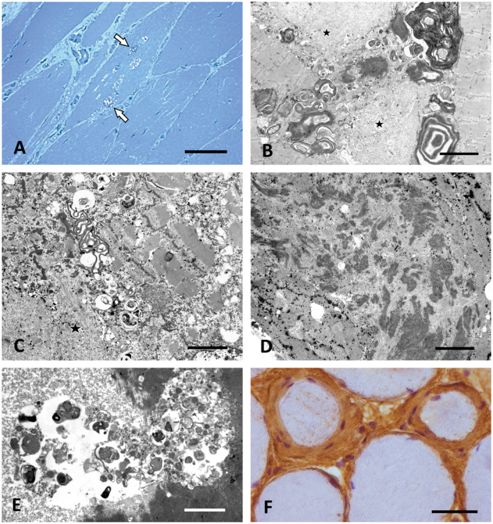
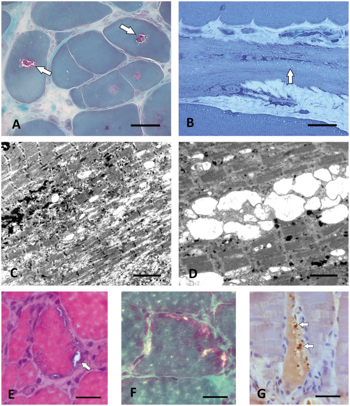
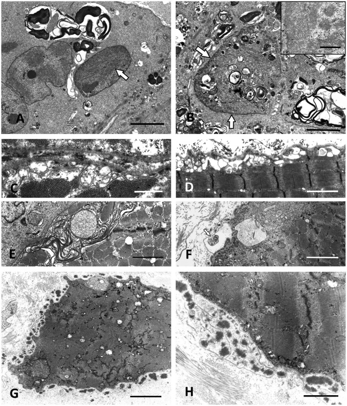
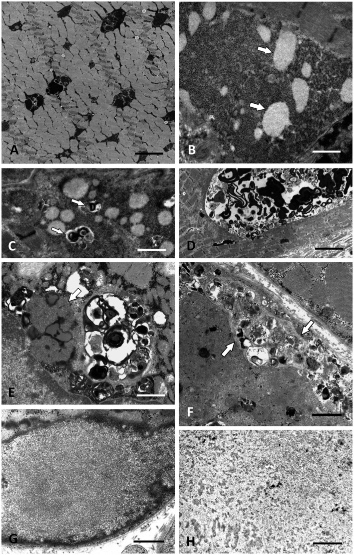
Altered autophagy accompanied by abnormal autophagic (rimmed) vacuoles detectable by light and electron microscopy is a common denominator of many familial and sporadic non-inflammatory muscle diseases. Even in the era of next generation sequencing (NGS), late-onset vacuolar myopathies remain a diagnostic challenge. We identified 32 adult vacuolar myopathy patients from 30 unrelated families, studied their clinical, histopathological and ultrastructural characteristics and performed genetic testing in index patients and relatives using Sanger sequencing and NGS including whole exome sequencing (WES). We established a molecular genetic diagnosis in 17 patients. Pathogenic mutations were found in genes typically linked to vacuolar myopathy (GNE, LDB3/ZASP, MYOT, DES and GAA), but also in genes not regularly associated with severely altered autophagy (FKRP, DYSF, CAV3, COL6A2, GYG1 and TRIM32) and in the digenic facioscapulohumeral muscular dystrophy 2. Characteristic histopathological features including distinct patterns of myofibrillar disarray and evidence of exocytosis proved to be helpful to distinguish causes of vacuolar myopathies. Biopsy validated the pathogenicity of the novel mutations p.(Phe55*) and p.(Arg216*) in GYG1 and of the p.(Leu156Pro) TRIM32 mutation combined with compound heterozygous deletion of exon 2 of TRIM32 and expanded the phenotype of Ala93Thr-caveolinopathy and of limb-girdle muscular dystrophy 2i caused by FKRP mutation. In 15 patients no causal variants were detected by Sanger sequencing and NGS panel analysis. In 12 of these cases, WES was performed, but did not yield any definite mutation or likely candidate gene. In one of these patients with a family history of muscle weakness, the vacuolar myopathy was eventually linked to chloroquine therapy. Our study illustrates the wide phenotypic and genotypic heterogeneity of vacuolar myopathies and validates the role of histopathology in assessing the pathogenicity of novel mutations detected by NGS. In a sizable portion of vacuolar myopathy cases, it remains to be shown whether the cause is hereditary or degenerative.

Keywords: FSHD; Pompe disease; TRIM32; autophagy; glycogenin 1; muscular dystrophy; myofibrillar myopathy; next generation sequencing (NGS); sarcotubular myopathy; vacuolar myopathy.


Figures

Figure 1
Figure 1
Characteristic morphological findings in vacuolar myopathies. A. Prominent vacuoles containing granular osmiophilic material (arrows) in GNE myopathy (P14). Semithin section, toluidine blue. Scale bar = 40 µm. B,C. Large osmiophilic membranous and granular inclusions indicative of altered autophagy combined with granulofilamentous material and other myofibrillar breakdown products (asterisks) in desminopathy (P7). EM. Scale bar in B = 3 µm, in C = 1.5 µm. D. Filamentous bundles characteristic for ZASPopathy in P10. EM. Scale bar = 3 µm. E. Large autophagic vacuole containing pleomorphic granular and membranous material in a case of Bethlem myopathy (P11) caused by COL6A2 mutation. EM. Scale bar = 2 µm. F. Prominent endomysial fibrosis with concentric accumulation of collagen and of fibroblasts around individual muscle fibers in P11. Paraffin section, Coll VI immunohistochemistry (brown). Scale bar = 30 µm.
Figure 2
Figure 2
Rimmed vacuoles in sarcotubular myopathy and FSHD2. A. Central rimmed vacuoles (white arrows) in P1.2 with compound heterozygous TRIM32 mutations. Cryostat section, Gömöri trichrome. Scale bar = 30 µm. B. Numerous smaller empty vacuoles arranged in rows typical for sarcotubular myopathy in P1.1, the brother of P1.2 who carried the same compound heterozygous TRIM32 mutations as P1.2. Semithin section, toluidine blue; scale bar = 30 µm. C,D. By EM, the small vacuoles in the biopsy of P1.1. At least partially correspond to dilations of the sarcoplasmic reticulum. Scale bar in C = 4 µm, in D = 2 µm. E. Muscle biopsy of the FSHD2 patient P6 showing subsarcolemmal rimmed vacuoles (arrow). Cryostat section, H&E. Scale bar = 20 µm. F. Subsarcolemmal rimmed vacuoles in the same biopsy as in (E). Cryostat section, Gömöri trichrome. Scale bar = 20 µm. G. The autophagic vacuoles in P6 contain pTDP‐43‐immunoreactive granular material (arrows). Paraffin section. Scale bar = 30 µm.
Figure 3
Figure 3
Ultrastructural pathology of FKRP, CAV3 and DYSF mutation cases. A. Prominent autophagic vacuoles combined with intranuclear tubulofilamentous inclusions (white arrow) in LGMD2i (LGMDR9) caused by compound heterozygous FKRP mutations (P3). Scale bar = 3 µm. B. P3: Membranous autophagic material accumulating within a myonucleus (arrows) and in the perinuclear sarcoplasm. Scale bar = 3 µm. Inset: High power view of sarcoplasmic tubulofilaments. Scale bar = 0.5 µm. C,D. Subsarcolemmal vacuoles and proliferations of membranes in a case with CAV 3 mutation (P15). Scale bar in C = 1 µm and D = 2 µm. E. Similar subsarcolemmal membranous proliferations in LGMD2B caused by compound heterozygous DYSF mutations (P5.2). Scale bar = 2 µm. F. Exocytosis of membranous autophagic material in the dysferlinopathy patient P5.1, the sister of P5.2. Scale bar = 2 µm. G,H. Large extracellular electron‐dense globules with abundant amorphous extracellular material surrounded by surplus basal lamina in the biopsy of P5.1. Scale bar in G = 10 µm, in H = 1.5 µm.
Figure 4 1
Figure 4 1
Ultrastructure of GSD II, GSD XV, chloroquine myopathy and cases without identifiable gene defect. A. Intermyofibrillar deposits of excess glycogen in GSD II (Pompe disease; P12). Scale bar = 3 µm. B. Muscle biopsy of P13 with novel compound heterozygous GYG1 (GSD XV) mutations showing excess glycogen with distinct round clusters of less osmiophilic fine granules, possibly representing partially degraded glycogen (arrows). Scale bar = 2 µm. C. P13: Autophagic vacuoles (arrows) associated with the glycogen deposits. Scale bar = 2.5 µm. D. P13: Large intermyofibrillar autophagic vacuole. Scale bar = 8 µm. E. Autophagic vacuoles combined with extensive accumulations of lipofuscin and curvilinear material (arrow) typical for chloroquine myopathy (P16). Scale bar = 2 µm. F. Arrows marking the membranous borders of exocytosed material between the plasma membrane and the basal lamina in P21. However, no mutation in LAMP‐2 or any other myopathy gene was found in this patient by WES. Scale bar = 2 µm. G. Distinct OPMD‐like tubulofilaments within a myonucleus in P27, who did not harbor a detectable PABPN1 repeat expansion or any other mutation in a myopathy gene. Scale bar = 0.5 µm. H. Deposition of granulofilamentous material in P19 suggested the diagnosis of a desminopathy/myofibrillar myopathy, but no defect in any of the relevant known genes was detected by WES. Scale bar = 0.5 µm.

References

    1. Al‐Tahan S, Weiss L, Yu H, Tang S, Saporta M, Vihola A et al (2019) New family with HSPB8‐associated autosomal dominant rimmed vacuolar myopathy. Neurol Genet 5:e349. - PMC - PubMed
    1. Amburgey K, Bailey A, Hwang JH, Tarnopolsky MA, Bonnemann CG, Medne L et al (2013) Genotype‐phenotype correlations in recessive RYR1‐related myopathies. Orphanet J Rare Dis 8:1–12. - PMC - PubMed
    1. Atsumi T, Ishikawa S, Miyatake T, Yoshida M (1979) Myopathy and primary aldosteronism: electronmicroscopic study. Neurology 29:1348–1353. - PubMed
    1. Berardo A, Lornage X, Johari M, Evangelista T, Cejas C, Barroso F et al (2019) HNRNPDL‐related muscular dystrophy: expanding the clinical, morphological and MRI phenotypes. J Neurol 266:2524–2534. - PubMed
    1. Brauers E, Dreier A, Roos A, Wormland B, Weis J, Krüttgen A (2010) Differential effects of myopathy‐associated caveolin‐3 mutants on growth factor signaling. Am J Pathol 177:261–270. - PMC - PubMed

Publication types

Supplementary concepts