

Simultaneous Nanoscale Imaging of Chemical and Architectural Heterogeneity on Yeast Cell Wall Particles
- PMID: 32419466
- PMCID: PMC7882198
- DOI: 10.1021/acs.langmuir.0c00627
Simultaneous Nanoscale Imaging of Chemical and Architectural Heterogeneity on Yeast Cell Wall Particles
Abstract
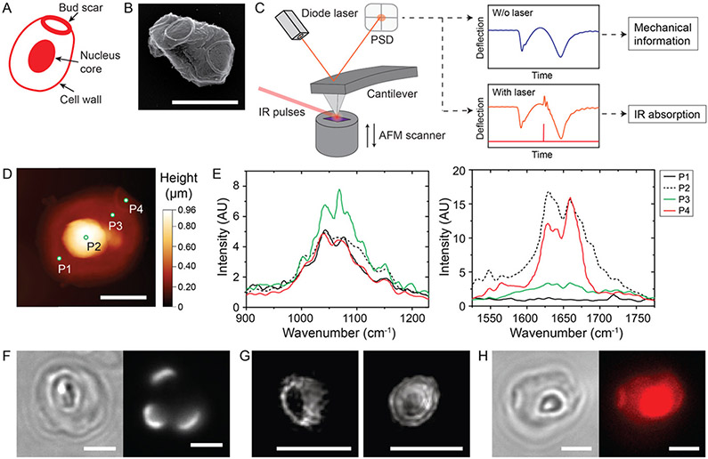
Particles extracted from yeast cell walls are naturally occurring immunomodulators with significant therapeutic applications. Their biological function has been thought to be a consequence of the overall chemical composition. In contrast, here we achieve direct nanoscale visualization of the compositional and structural heterogeneity of yeast cell wall particles and demonstrate that such nanoscale heterogeneity directly influences the receptor function of immune cells. By combining peak force infrared (PFIR) microscopy with super-resolution fluorescence microscopy, we achieve simultaneous chemical, topographical, and mechanical mapping of cell wall particles extracted from the yeast Saccharomyces cerevisiae with ≈6 nm resolution. We show that polysaccharides (β-glucan and chitin) and proteins are organized in specific nonuniform structures, and their heterogeneous spatial organization leads to heterogeneous recruitment of receptors on immune cell membranes. Our findings indicate that the biological function of yeast cell wall particles depends on not only their overall composition but also the nanoscale distribution of the different cell wall components.
Figures


Similar articles
-
Saccharomyces cerevisiae CellWall Remodeling in the Absence of Knr4 and Kre6 Revealed by Nano-FourierTransform Infrared Spectroscopy.Appl Spectrosc. 2024 Apr;78(4):355-364. doi: 10.1177/00037028231213658. Epub 2024 Feb 20. Appl Spectrosc. 2024. PMID: 38378014 Free PMC article.
-
Increase in chitin as an essential response to defects in assembly of cell wall polymers in the ggp1delta mutant of Saccharomyces cerevisiae.J Bacteriol. 1997 Jan;179(2):463-9. doi: 10.1128/jb.179.2.463-469.1997. J Bacteriol. 1997. PMID: 8990299 Free PMC article.
-
Production of yeast cell wall polysaccharides-β-glucan and chitin by using food waste substrates: Biosynthesis, production, extraction, and purification methods.Compr Rev Food Sci Food Saf. 2025 May;24(3):e70161. doi: 10.1111/1541-4337.70161. Compr Rev Food Sci Food Saf. 2025. PMID: 40183630 Free PMC article. Review.
-
Extraction of two active polysaccharides from the yeast cell wall.Z Naturforsch C J Biosci. 2008 Nov-Dec;63(11-12):919-21. doi: 10.1515/znc-2008-11-1224. Z Naturforsch C J Biosci. 2008. PMID: 19227846
-
Cell wall polysaccharides: before and after autolysis of brewer's yeast.World J Microbiol Biotechnol. 2018 Aug 20;34(9):137. doi: 10.1007/s11274-018-2508-6. World J Microbiol Biotechnol. 2018. PMID: 30128783 Review.
Cited by
-
Liquid-Phase Peak Force Infrared Microscopy for Chemical Nanoimaging and Spectroscopy.Anal Chem. 2021 Feb 23;93(7):3567-3575. doi: 10.1021/acs.analchem.0c05075. Epub 2021 Feb 11. Anal Chem. 2021. PMID: 33573375 Free PMC article.
-
Nanoscale Chemical Features of the Natural Fibrous Material Wood.Biomacromolecules. 2020 Oct 12;21(10):4244-4252. doi: 10.1021/acs.biomac.0c01028. Epub 2020 Sep 11. Biomacromolecules. 2020. PMID: 32852940 Free PMC article.
-
AFM-IR for Nanoscale Chemical Characterization in Life Sciences: Recent Developments and Future Directions.ACS Meas Sci Au. 2023 Jun 16;3(5):301-314. doi: 10.1021/acsmeasuresciau.3c00010. eCollection 2023 Oct 18. ACS Meas Sci Au. 2023. PMID: 37868358 Free PMC article. Review.
-
An important polysaccharide from fermentum.Food Chem X. 2022 Jul 8;15:100388. doi: 10.1016/j.fochx.2022.100388. eCollection 2022 Oct 30. Food Chem X. 2022. PMID: 36211774 Free PMC article. Review.
-
Inducing trained immunity in pro-metastatic macrophages to control tumor metastasis.Nat Immunol. 2023 Feb;24(2):239-254. doi: 10.1038/s41590-022-01388-8. Epub 2023 Jan 5. Nat Immunol. 2023. PMID: 36604547 Free PMC article.
References
-
- Novak M; Vetvicka V B-Glucans, History, and the Present: Immunomodulatory Aspects and Mechanisms of Action. J. Immunotoxicol 2008, 5, 47–57. - PubMed
-
- De Smet R; Demoor T; Verschuere S; Dullaers M; Ostroff GR; Leclercq G; Allais L; Pilette C; Dierendonck M; De Geest BG; Cuvelier CA B-Glucan Microparticles are Good Candidates for Mucosal Antigen Delivery in Oral Vaccination. J. Controlled Release 2013, 172, 671–678. - PubMed
Publication types
MeSH terms
Substances
Grants and funding
LinkOut - more resources
Full Text Sources
Molecular Biology Databases
