

Treatment Strategies for Tandem Occlusions in Acute Ischemic Stroke
- PMID: 32419734
- PMCID: PMC7224971
- DOI: 10.1055/s-0040-1709207
Treatment Strategies for Tandem Occlusions in Acute Ischemic Stroke
Abstract
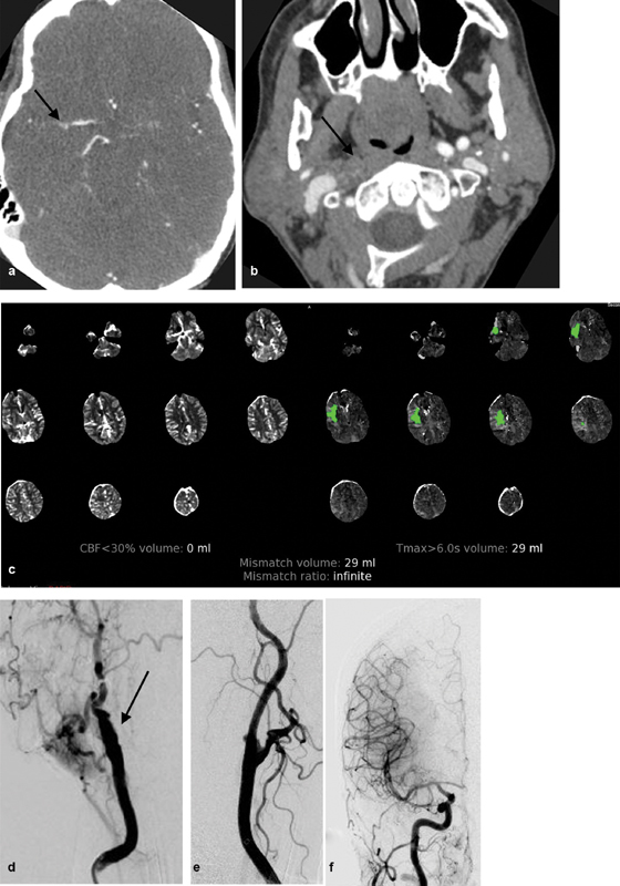
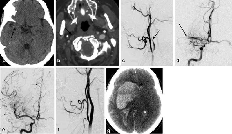
There is no consensus for the treatment of a tandem occlusion (TO) in a patient presenting with an acute ischemic stroke. In this review article, we will focus on the controversial treatment strategies for TOs. First, we will discuss treatment options including retrograde, antegrade, and delayed approaches. Second, the role of carotid stent placement versus balloon angioplasty for the extracranial occlusion will be presented. Third, anticoagulation and antiplatelet regimens for the treatment TOs published in the literature will be reviewed. Finally, we will discuss whether there is a role for coil occlusion of the cervical carotid artery or whether staged carotid revascularization days after mechanical thrombectomy of the intracranial occlusion maybe appropriate. The optimal treatment strategy of TO has not been established and further larger trials need to be performed to answer the question.
Keywords: angioplasty; embolization; interventional radiology; occlusion; stenosis; stent; stroke.
© Thieme Medical Publishers.
Conflict of interest statement
Conflict of Interest None declared.
Figures
Similar articles
-
Diagnosis and management of tandem occlusion in acute ischemic stroke.Eur J Radiol Open. 2023 Aug 14;11:100513. doi: 10.1016/j.ejro.2023.100513. eCollection 2023 Dec. Eur J Radiol Open. 2023. PMID: 37609048 Free PMC article.
-
Acute Ischemic Stroke due to Common Carotid Ostial Disease with Tandem Intracranial Occlusions Treated with Thrombectomy and Staged Retrograde Stenting.Interv Neurol. 2018 Oct;7(6):445-451. doi: 10.1159/000490584. Epub 2018 Jul 13. Interv Neurol. 2018. PMID: 30410523 Free PMC article.
-
Revascularization of tandem occlusions in acute ischemic stroke: review of the literature and illustrative case.Neurosurg Focus. 2017 Apr;42(4):E15. doi: 10.3171/2017.1.FOCUS16521. Neurosurg Focus. 2017. PMID: 28366063 Review.
-
Simultaneous revascularization of the occluded internal carotid artery using the Solitaire as a workhorse wire during acute ischemic stroke intervention.Interv Neuroradiol. 2020 Apr;26(2):205-210. doi: 10.1177/1591019919885253. Epub 2019 Nov 7. Interv Neuroradiol. 2020. PMID: 31696768 Free PMC article.
-
Overview of evidence on emergency carotid stenting in patients with acute ischemic stroke due to tandem occlusions: a systematic review and meta-analysis.J Cardiovasc Surg (Torino). 2019 Dec;60(6):693-702. doi: 10.23736/S0021-9509.18.10312-0. Epub 2018 Jan 23. J Cardiovasc Surg (Torino). 2019. PMID: 29363895
Cited by
-
Predictors of outcome after endovascular treatment for tandem occlusions: a single center retrospective analysis.BMC Neurol. 2023 Feb 27;23(1):82. doi: 10.1186/s12883-023-03127-4. BMC Neurol. 2023. PMID: 36849925 Free PMC article.
References
-
- Rubiera M, Ribo M, Delgado-Mederos R et al.Tandem internal carotid artery/middle cerebral artery occlusion: an independent predictor of poor outcome after systemic thrombolysis. Stroke. 2006;37(09):2301–2305. - PubMed
-
- Kim Y S, Garami Z, Mikulik R, Molina C A, Alexandrov A V; CLOTBUST Collaborators.Early recanalization rates and clinical outcomes in patients with tandem internal carotid artery/middle cerebral artery occlusion and isolated middle cerebral artery occlusion Stroke 20053604869–871. - PubMed
-
- National Institute of Neurological Disorders and Stroke rt-PA Stroke Study Group.Tissue plasminogen activator for acute ischemic stroke N Engl J Med 1995333241581–1587. - PubMed
-
- Christou I, Felberg R A, Demchuk A M et al.Intravenous tissue plasminogen activator and flow improvement in acute ischemic stroke patients with internal carotid artery occlusion. J Neuroimaging. 2002;12(02):119–123. - PubMed
-
- Rubiera M, Alvarez-Sabín J, Ribo M et al.Predictors of early arterial reocclusion after tissue plasminogen activator-induced recanalization in acute ischemic stroke. Stroke. 2005;36(07):1452–1456. - PubMed
