

Effect of Electroacupuncture in Mice with Dextran Sulfate Sodium-Induced Colitis and the Influence of Gut Microbiota
- PMID: 32419794
- PMCID: PMC7204379
- DOI: 10.1155/2020/2087903
Effect of Electroacupuncture in Mice with Dextran Sulfate Sodium-Induced Colitis and the Influence of Gut Microbiota
Abstract
Background: The relationship between inflammatory bowel disease and gut microbiota is inextricable. Electroacupuncture (EA) can alleviate acute experimental colitis, but the performance of intestinal microorganisms and the mechanism are still not fully understood. We investigated the relationship between the EA and gut microbes and clarified the role of tight junction and adiponectin in the anti-inflammatory effect of EA.
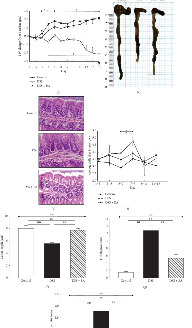
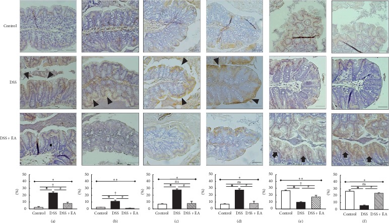
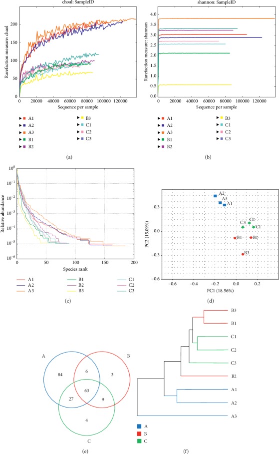
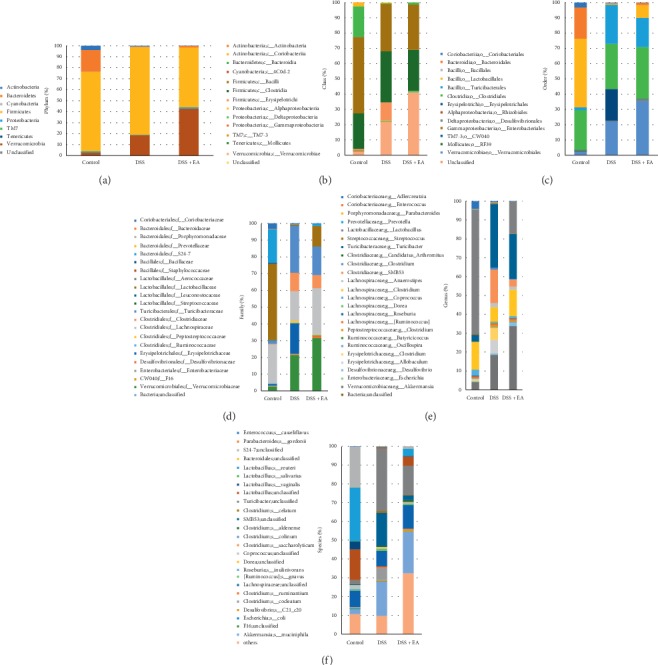
Methods: Male C57BL/6 mice were randomized into three groups: normal control, dextran sulfate sodium- (DSS-) induced ulcerative colitis (DSS), and DSS with EA ST36 (DSS + EA). Mice body weight, DAI score, colon length, and histological score were evaluated for colitis severity. Colonic inflammation and tight junctions were demonstrated by the immunohistochemical (IHC) method. Systemic responses were confirmed by plasma cytokines and adiponectin with multiplex immunoassays. Gut microbiome profiling was conducted by 16S rRNA gene sequencing.
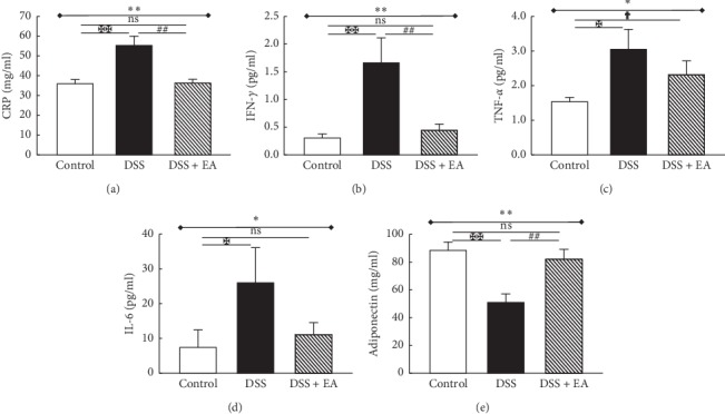
Results: EA had benefit in relieving both macroscopic and microscopic colonic inflammation. It can reduce disease activity, maintain colon length, and ameliorate histological inflammatory reaction. In IHC stain, EA decreased CD11b, F4/80, TLR4, and MyD88 and preserved claudin-1 and ZO-1 expression. Compared with the control group, the DSS group showed elevated levels of CRP, IFN-γ, TNF-α, and IL-6, but decreased adiponectin. These changes were reversed by EA, accompanied by modulation of the overall structure of gut microbiota.
Conclusion: Our findings suggest that EA exerts its therapeutic effect by TLR4 signaling via the MyD88-dependent pathway. EA could increase adiponectin, maintain mucosal tight junctions, and modulate gut microbiota.
Copyright © 2020 Geng-Hao Liu et al.
Conflict of interest statement
The authors declare that they have no conflicts of interest.
Figures
Similar articles
-
Electroacupuncture preserves intestinal barrier integrity through modulating the gut microbiota in DSS-induced chronic colitis.Life Sci. 2020 Nov 15;261:118473. doi: 10.1016/j.lfs.2020.118473. Epub 2020 Sep 21. Life Sci. 2020. PMID: 32971101
-
Alterations in Gut Microbiota and Upregulations of VPAC2 and Intestinal Tight Junctions Correlate with Anti-Inflammatory Effects of Electroacupuncture in Colitis Mice with Sleep Fragmentation.Biology (Basel). 2022 Jun 25;11(7):962. doi: 10.3390/biology11070962. Biology (Basel). 2022. PMID: 36101343 Free PMC article.
-
2,3,5,4'-Tetrahydroxystilbene-2-O-β-D-glucoside, a major bioactive component from Polygoni multiflori Radix (Heshouwu) suppresses DSS induced acute colitis in BALb/c mice by modulating gut microbiota.Biomed Pharmacother. 2021 May;137:111420. doi: 10.1016/j.biopha.2021.111420. Epub 2021 Feb 23. Biomed Pharmacother. 2021. PMID: 33761623
-
Electroacupuncture repairs intestinal barrier by upregulating CB1 through gut microbiota in DSS-induced acute colitis.Chin Med. 2023 Mar 10;18(1):24. doi: 10.1186/s13020-023-00733-9. Chin Med. 2023. PMID: 36894930 Free PMC article.
-
Modulation of Gut Microbiota Combined with Upregulation of Intestinal Tight Junction Explains Anti-Inflammatory Effect of Corylin on Colitis-Associated Cancer in Mice.Int J Mol Sci. 2022 Feb 28;23(5):2667. doi: 10.3390/ijms23052667. Int J Mol Sci. 2022. PMID: 35269806 Free PMC article.
Cited by
-
Electroacupuncture alleviates ulcerative colitis by targeting CXCL1: evidence from the transcriptome and validation.Front Immunol. 2023 Sep 1;14:1187574. doi: 10.3389/fimmu.2023.1187574. eCollection 2023. Front Immunol. 2023. PMID: 37727787 Free PMC article.
-
Acupuncture influences multiple diseases by regulating gut microbiota.Front Cell Infect Microbiol. 2024 Jul 8;14:1371543. doi: 10.3389/fcimb.2024.1371543. eCollection 2024. Front Cell Infect Microbiol. 2024. PMID: 39040602 Free PMC article. Review.
-
Matrix metalloproteinases are key targets of acupuncture in the treatment of ulcerative colitis.Exp Biol Med (Maywood). 2023 Jul;248(14):1229-1241. doi: 10.1177/15353702231182205. Epub 2023 Jul 12. Exp Biol Med (Maywood). 2023. PMID: 37438919 Free PMC article.
-
Slight Disruption in Intestinal Environment by Dextran Sodium Sulfate Reduces Egg Yolk Size Through Disfunction of Ovarian Follicle Growth.Front Physiol. 2021 Jan 15;11:607369. doi: 10.3389/fphys.2020.607369. eCollection 2020. Front Physiol. 2021. PMID: 33519513 Free PMC article.
-
Acupuncture and moxibustion therapy for cognitive impairment: the microbiome-gut-brain axis and its role.Front Neurosci. 2024 Jan 11;17:1275860. doi: 10.3389/fnins.2023.1275860. eCollection 2023. Front Neurosci. 2024. PMID: 38274501 Free PMC article. Review.
References
LinkOut - more resources
Full Text Sources
Research Materials
Miscellaneous
