

Screening CT angiography in patients with suspected fibromuscular dysplasia: improved patient care with single-session skull vertex to pelvis coverage
- PMID: 32420100
- PMCID: PMC7225442
- DOI: 10.21037/cdt.2020.02.06
Screening CT angiography in patients with suspected fibromuscular dysplasia: improved patient care with single-session skull vertex to pelvis coverage
Abstract
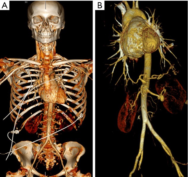
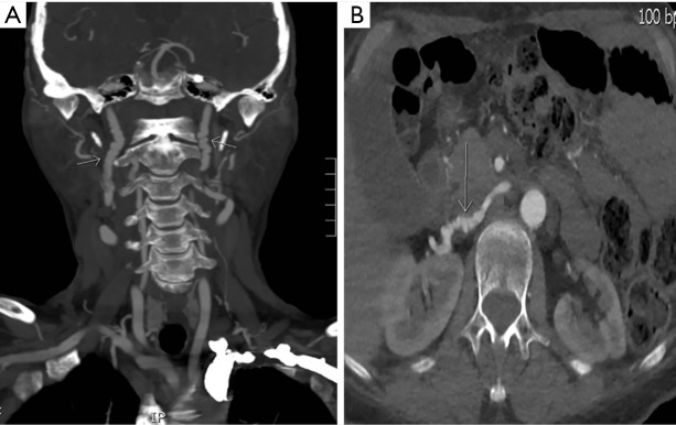
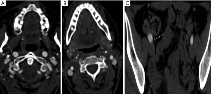
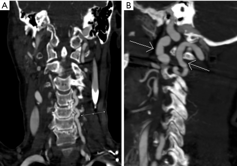
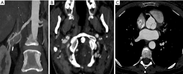
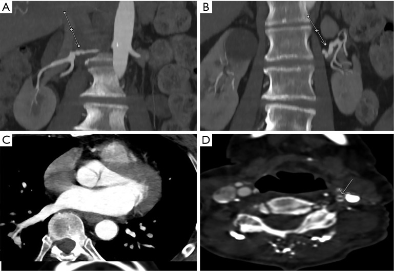
Background: Imaging plays a key role in the workup of patients with clinically suspected fibromuscular dysplasia (FMD), and research has highlighted the potential of computed tomography angiography (CTA) in screening for thoracic, abdominal, and pelvic arterial abnormalities in these patients. We sought to evaluate imaging findings from patients with suspected or diagnosed FMD who underwent screening CTA at our institution with a novel single-acquisition protocol that offers increased anatomic coverage, with images obtained from the skull vertex to the pelvis.
Methods: Images from 80 consecutive patients scanned with the novel single-session CTA protocol covering the skull vertex to the pelvis were compared with images from 20 additional consecutive patients who underwent CTA for the head and neck separate from CTA of the chest, abdomen, and pelvis.
Results: Compared with CTA performed in separate sessions, the single-session CTA protocol decreased the radiation dose by 38% (P<0.001) and decreased the contrast dose by 39% (P<0.001), with satisfactory image quality noted in all instances. Additionally, higher mean contrast attenuation was noted in the aortic arch with use of the novel protocol (409±76 HU) versus with use of the dual-acquisition protocol (260±38 HU; P<0.001).
Conclusions: These results suggest that use of a novel single-session CTA protocol extending from the skull vertex to the pelvis provides effective screening imaging in patients with suspected or diagnosed FMD as compared with multisession, standard-pitch CTA.
Keywords: Computed tomography angiography (CTA); fibromuscular dysplasia (FMD); screening.
2020 Cardiovascular Diagnosis and Therapy. All rights reserved.
Conflict of interest statement
Conflicts of Interest: Both authors have completed the ICMJE uniform disclosure form (available at http://dx.doi.org/10.21037/cdt.2020.02.06). The authors have no conflicts of interest to declare.
Figures
References
LinkOut - more resources
Full Text Sources