

The influence of smoking exposure and cessation on penile hemodynamics and corporal tissue in a rat model
- PMID: 32420170
- PMCID: PMC7215033
- DOI: 10.21037/tau.2019.12.45
The influence of smoking exposure and cessation on penile hemodynamics and corporal tissue in a rat model
Abstract
Background: While epidemiological studies have clearly documented that smoking cessation significantly enhances sexual health, the underlying mechanism remains largely unknown. Thus, we wished to explore possible mechanisms by using a rat model of smoking-associated erectile dysfunction (ED).
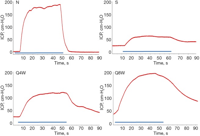
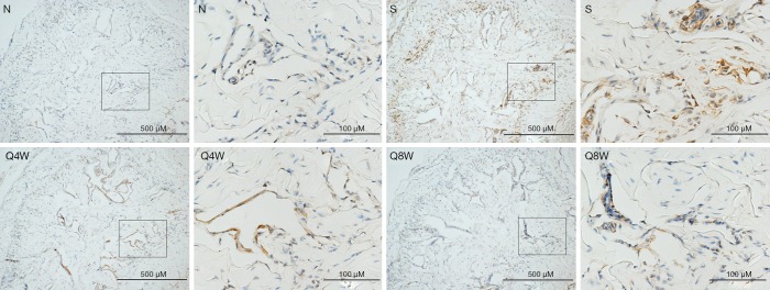
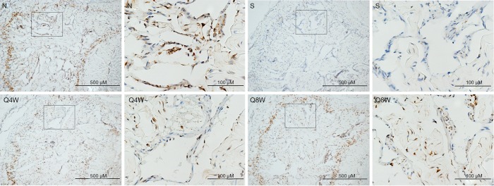
Methods: Forty 8-week old male Sprague-Dawley rats were divided into 4 groups. Ten rats were exposed only to room air (N group). The remaining 30 rats were passively exposed to cigarette smoke over a 12-week period. At the end of 12 weeks, the smoking (S, n=10) group underwent immediate erectile function testing and were sacrificed. The remaining 20 rats were exposed to room air only for 4 (Q4W, n=10) or 8 (Q8W, n=10) weeks and then underwent erectile function testing and sacrifice. Erectile function was evaluated by measuring intracavernous pressure (ICP) and mean arterial pressure (MAP). After blood collection for serum testosterone determination, rats were sacrificed to obtain corporal tissue for immunohistochemistry.
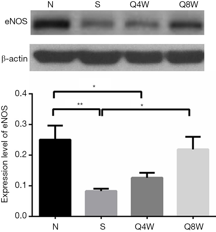
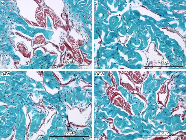
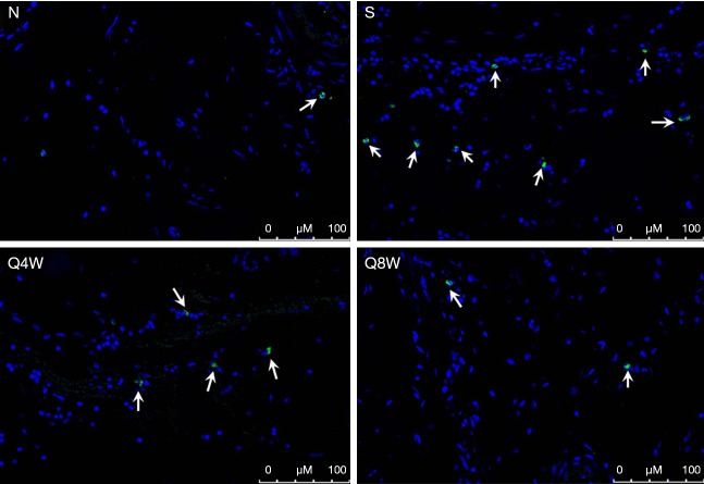
Results: Mean ICP/MAP ratio was significantly lower in the S group compared to the N and Q8W groups (0.52±0.11, 0.94±0.05, and 0.94±0.12, respectively, P=0.0189). Smooth muscle/collagen ratio was also significantly lower in the S group compared to the N and Q8W groups (11.8±0.94, 17.5±1.82, and 16.4±0.60, respectively, P=0.0008). Oxidative stress and apoptotic indices were significantly higher in the S group compared to the N and Q8W groups. Neuronal and endothelial nitric oxide synthases were significantly less expressed in the S group compared to the N and Q8W groups.
Conclusions: Smoking cessation is associated with partial recovery of penile hemodynamics in a rat model of smoking associated ED.
Keywords: Cigarette; cessation; erectile dysfunction (ED); rat; smoking.
2020 Translational Andrology and Urology. All rights reserved.
Conflict of interest statement
Conflicts of Interest: All authors have completed the ICMJE uniform disclosure form (available at http://dx.doi.org/10.21037/tau.2019.12.45). The authors have no conflicts of interest to declare.
Figures
Similar articles
-
Chronic Cigarette Smoking Impairs Erectile Function through Increased Oxidative Stress and Apoptosis, Decreased nNOS, Endothelial and Smooth Muscle Contents in a Rat Model.PLoS One. 2015 Oct 22;10(10):e0140728. doi: 10.1371/journal.pone.0140728. eCollection 2015. PLoS One. 2015. PMID: 26491965 Free PMC article.
-
The Effects of Adipose-Derived Stem Cells in a Rat Model of Tobacco-Associated Erectile Dysfunction.PLoS One. 2016 Jun 3;11(6):e0156725. doi: 10.1371/journal.pone.0156725. eCollection 2016. PLoS One. 2016. PMID: 27257818 Free PMC article.
-
Dietary Modification Is Associated with Normalization of Penile Hemodynamics in Rats Fed a High-Fat Diet.J Sex Med. 2019 Jun;16(6):791-802. doi: 10.1016/j.jsxm.2019.03.013. Epub 2019 Apr 19. J Sex Med. 2019. PMID: 31010783
-
Chronic prostatitis/chronic pelvic pain syndrome impairs erectile function through increased endothelial dysfunction, oxidative stress, apoptosis, and corporal fibrosis in a rat model.Andrology. 2016 Nov;4(6):1209-1216. doi: 10.1111/andr.12273. Epub 2016 Aug 27. Andrology. 2016. PMID: 27565759
-
Intracavernous Injection of Autologous Platelet-Rich Plasma Ameliorates Hyperlipidemia-Associated Erectile Dysfunction in a Rat Model.Sex Med. 2021 Apr;9(2):100317. doi: 10.1016/j.esxm.2020.100317. Epub 2021 Jan 30. Sex Med. 2021. PMID: 33529811 Free PMC article.
Cited by
-
Erectile Dysfunction and Oxidative Stress: A Narrative Review.Int J Mol Sci. 2025 Mar 27;26(7):3073. doi: 10.3390/ijms26073073. Int J Mol Sci. 2025. PMID: 40243750 Free PMC article. Review.
-
Oxidative Stress and Erectile Dysfunction: Pathophysiology, Impacts, and Potential Treatments.Curr Issues Mol Biol. 2024 Aug 14;46(8):8807-8834. doi: 10.3390/cimb46080521. Curr Issues Mol Biol. 2024. PMID: 39194738 Free PMC article. Review.
References
LinkOut - more resources
Full Text Sources