Case Reports
. 2020 Apr;9(2):794-799.
doi: 10.21037/tau.2020.02.06.

Case report: IgG4-related renal disease co-existing with retroperitoneal fibrosis

Affiliations
Case Reports

Case report: IgG4-related renal disease co-existing with retroperitoneal fibrosis

Young Jin Kim et al. Transl Androl Urol. 2020 Apr.

Abstract

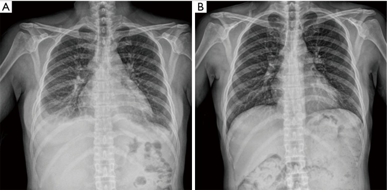
A 51-year-old woman visited our clinic for orthopnea, chest discomfort, and facial and leg edema. Acute kidney injury and pleural effusion were diagnosed and she received hemodialysis. This is a rare case of IgG4-related disease diagnosed by renal biopsy in the patient with retroperitoneal fibrosis and severe acute kidney injury, which progressed rapidly and required hemodialysis without markedly elevated serum IgG4 level and urinary tract obstruction. Immunostaining of a kidney biopsy revealed IgG4-related renal disease and magnetic resonance imaging showed retroperitoneal fibrosis in the paraspinal and presacral areas. After 3 days of glucocorticoid treatment, renal function and bilateral pleural effusion were restored. Another 2 months of glucocorticoid treatment decreased the extent of retroperitoneal fibrosis on follow-up abdominal computed tomography. IgG4-related disease is a recently recognized fibro-inflammatory condition with a dense lymphoplasmacytic infiltration of IgG4-positive plasma cells. The kidney and retroperitoneum are the frequently associated organs in IgG4-related disease. This case shows that IgG4-related disease can involve the kidney and/or other organs and be diagnosed by kidney biopsy.

Keywords: IgG4-related renal disease; acute kidney injury; case report; kidney biopsy; retroperitoneal fibrosis.


Conflict of interest statement

Conflict of interest: All authors have completed the ICMJE uniform disclosure form (available at http://dx.doi.org/10.21037/tau.2020.02.06). The authors have no conflicts of interest to declare.

Figures

Figure 1
Figure 1
Chest X-rays before and after treatment. (A) Bilateral pleural effusion and pulmonary edema on chest simple X-ray. (B) Pleural effusion and pulmonary edema was disappeared after treatment.
Figure 2
Figure 2
Kidney ultrasonography. Kidney ultrasonography showing increased cortical echogenicity with mild bilateral hydronephrosis (grade 1).
Figure 3
Figure 3
Kidney biopsy. A tissue specimen form kidney biopsy showed a dense lymphoplasmacytic infiltration (A, hematoxylin and eosin stain, ×200; B, Periodic acid-Schiff stain, ×200; C, Masson trichrome stain, ×100). Immunohistochemical staining for IgG4 monoclonal antibody showed more than 10 IgG4-positive plasma cells per high-power field in the peritubular interstitial area (D, ×200).
Figure 4
Figure 4
Positron emission tomography-computed tomography (PET-CT). PET-CT with 18fluorodeoxyglucose revealed a diffuse hypermetabolic soft tissue lesion along the retroperitoneal space (arrow). There was no significant hypermetabolism in the lung and pleura.
Figure 5
Figure 5
Clinical course of IgG4 related renal disease. HD, hemodialysis; Bx., Kidney biopsy; PRD, prednisolone; CRP, C-reactive peptide.
Figure 6
Figure 6
Enhanced CT scans of the abdomen, before and after treatment. Enhanced CT scan of the abdomen revealed diffuse prevertebral soft tissue thickening and enhancement at lower lumbar spine and sacrum (A, arrow). The CT scan after treatment showed a decreased extent of the ill-defined soft tissue lesion involving both the paraspinal area of L5 and the presacral space (B, arrow).

Similar articles

Cited by

References

    1. Stone JH, Zen Y, Deshpande V. IgG4-related disease. N Engl J Med 2012;366:539-51. 10.1056/NEJMra1104650 - DOI - PubMed
    1. Ormond JK. Bilateral ureteral obstruction due to envelopment and compression by an inflammatory retroperitoneal process. J Urol 1948;59:1072-9. 10.1016/S0022-5347(17)69482-5 - DOI - PubMed
    1. Cronin CG, Lohan D, Blake M, et al. Retroperitoneal fibrosis: a review of clinical features and imaging findings. AJR Am J Roentgenol 2008;191:423-31. 10.2214/AJR.07.3629 - DOI - PubMed
    1. Cheuk W, Chan JK. IgG4-related sclerosing disease: a critical appraisal of an evolving clinicopathologic entity. Adv Anat Pathol 2010;17:303-32. 10.1097/PAP.0b013e3181ee63ce - DOI - PubMed
    1. Hamano H, Kawa S, Horiuchi A, et al. High serum IgG4 concentrations in patients with sclerosing pancreatitis. N Engl J Med 2001;344:732-8. 10.1056/NEJM200103083441005 - DOI - PubMed

Publication types