. 2020 Apr 25:2020:6708061.
doi: 10.1155/2020/6708061. eCollection 2020.

Catalpol Attenuates Hepatic Steatosis by Regulating Lipid Metabolism via AMP-Activated Protein Kinase Activation

Affiliations

Catalpol Attenuates Hepatic Steatosis by Regulating Lipid Metabolism via AMP-Activated Protein Kinase Activation

Xiang Tian et al. Biomed Res Int. .

Abstract

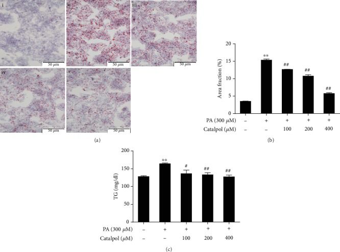
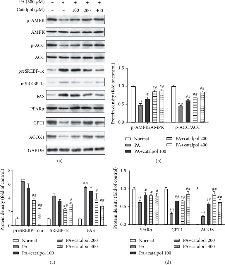
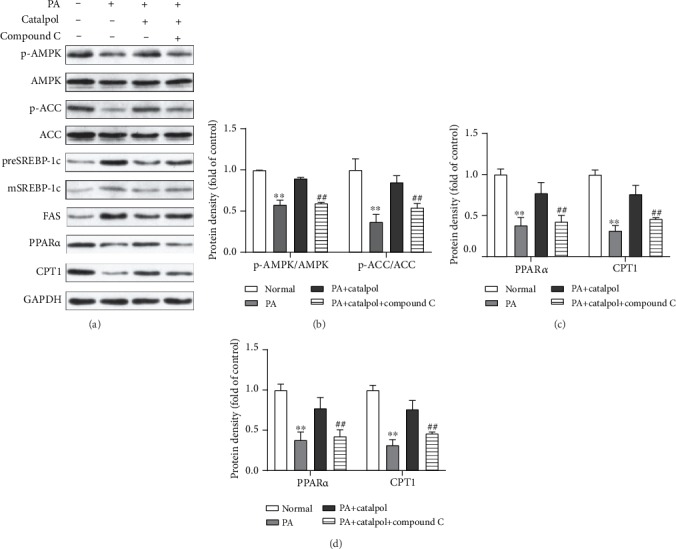
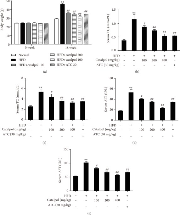
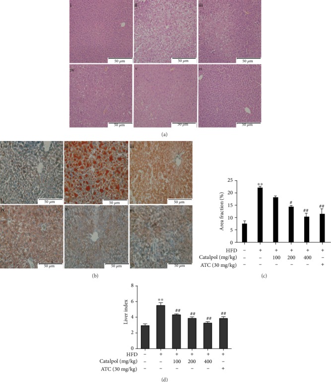
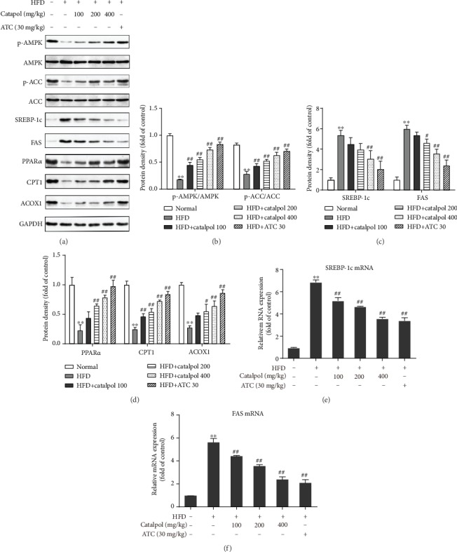
The increased prevalence of nonalcoholic fatty liver disease (NAFLD), which develops from hepatic steatosis, represents a public health challenge. Catalpol, a natural component extracted from the roots of Radix Rehmanniae, has several pharmacological activities. The present study is aimed at examining whether catalpol prevents hepatic steatosis in cell and animal experiments and elucidating the possible mechanisms. HepG2 cells were treated with 300 μM palmitate (PA) and/or catalpol for 24 h in vitro, and male C57BL/6J mice fed a high-fat diet (HFD) were administered catalpol for 18 weeks in vivo. The results revealed that catalpol significantly decreased lipid accumulation in PA-treated HepG2 cells. Moreover, catalpol drastically reduced body weight and lipid accumulation in the liver, whereas it ameliorated hepatocyte steatosis in HFD-fed mice. Notably, catalpol remarkably promoted the phosphorylation of AMP-activated protein kinase (AMPK) and acetyl-CoA carboxylase. Subsequently, catalpol repressed the expressions of lipogenesis-associated genes such as sterol regulatory element-binding protein 1c and fatty acid synthase but promoted the expressions of genes associated with fatty acid β-oxidation such as peroxisome proliferator-activated receptor α together with its target genes carnitine palmitoyltransferase 1 and acyl-CoA oxidase 1 (ACOX1). However, the preincubation of the HepG2 cells with compound C (10 μM), an AMPK inhibitor, prevented catalpol-mediated beneficial effects. These findings suggest that catalpol ameliorates hepatic steatosis by suppressing lipogenesis and enhancing fatty acid β-oxidation in an AMPK-dependent manner. Therefore, catalpol has potential as a novel agent in the treatment of NAFLD.


Conflict of interest statement

The authors have no conflict of interest to declare.

Figures

Figure 1
Figure 1
Catalpol inhibits palmitate- (PA-) induced triglyceride (TG) accumulation in HepG2 cells. HepG2 cells were treated with PA (300 μM) and/or catalpol (100, 200, or 400 μM) for 24 h. (a) Lipid accumulation was determined via Oil Red O staining. Images of cells were photographed at 200x magnification: (i) control, (ii) PA (300 μM), (iii) PA (300 μM)+catalpol (100 μM), (iv) PA (300 μM)+catalpol (200 μM), and (v) PA (300 μM)+catalpol (400 μM). (b) A larger red area consisting of lipid droplets in HepG2 cells. (c) Measurement of intracellular TG content. Data are presented as the mean ± SE of three independent experiments. ∗∗P < 0.01 vs. the Normal group; #P < 0.05, ##P < 0.01 vs. the PA group.
Figure 2
Figure 2
Catalpol treatment regulates enzymes and genes involved in lipid metabolism in palmitate- (PA-) treated HepG2 cells. HepG2 cells were treated with PA (300 μM) and/or catalpol (100, 200, or 400 μM) for 24 h. (a) Protein expressions of p-AMP-activated protein kinase (AMPK), p-acetyl-CoA carboxylase (ACC), precursor and mature sterol regulatory element-binding protein 1c (preSREBP-1c and mSREBP-1c, respectively), fatty acid synthase (FAS), peroxisome proliferator-activated receptor α (PPARα), carnitine palmitoyltransferase 1 (CPT1), and acyl-CoA oxidase 1 (ACOX1) were analyzed via Western blotting. (b–d) Densitometric analyses of the band intensity ratios of p-AMPK/AMPK, p-ACC/ACC, preSREBP-1c, mSREBP-1c, FAS, PPARα, CPT1, and ACOX1. Data are presented as the mean ± SE of three independent experiments. ∗∗P < 0.01 vs. the Normal group; #P < 0.05, ##P < 0.01 vs. the PA group.
Figure 3
Figure 3
AMPK activation mediates catalpol-regulated lipid metabolism in palmitate- (PA-) treated HepG2 cells. HepG2 cells were treated with PA (300 μM) and/or catalpol (400 μM) for 24 h. Compound C (10 μM) was added 2 h prior to the cotreatment with PA and catalpol. (a) Protein expressions of p-AMP-activated protein kinase (AMPK), p-acetyl-CoA carboxylase (ACC), precursor and mature sterol regulatory element-binding protein 1c (preSREBP-1c and mSREBP-1c, respectively), fatty acid synthase (FAS), peroxisome proliferator-activated receptor α (PPARα), and carnitine palmitoyltransferase 1 (CPT1) were analyzed via Western blotting. (b–d) Densitometric analyses of the band intensity ratios of p-AMPK/AMPK, p-ACC/ACC, preSREBP-1c, mSREBP-1c, FAS, PPARα, and CPT1. Data are presented as the mean ± SE of three independent experiments. ∗∗P < 0.01 vs. the Normal group; #P < 0.05, ##P < 0.01 vs. the catalpol group.
Figure 4
Figure 4
Catalpol treatment reduces body weight gain and elevates the serum levels of lipids and hepatic enzymes in high-fat diet- (HFD-) fed mice. C57BL/6J mice were fed a normal diet or HFD and treated with saline, atorvastatin calcium (ATC), or different doses of catalpol daily for 18 weeks. (a) Body weight changes. (b–e) Serum levels of triglyceride (TG), total cholesterol (TC), alanine aminotransferase (ALT), and aspartate aminotransferase (AST). Data are presented as the mean ± SE (n = 8). ∗∗P < 0.01 vs. the Normal group; #P < 0.05, ##P < 0.01 vs. the HFD group.
Figure 5
Figure 5
Catalpol treatment prevented hepatic steatosis in high-fat diet- (HFD-) fed mice. (a) Hematoxylin and eosin staining of the hepatic sections (200x): (i) normal, (ii) HFD, (iii) HFD+catalpol (100 mg/kg, oral gavage), (iv) HFD+catalpol (200 mg/kg, oral gavage), (v) HFD+catalpol (400 mg/kg, oral gavage), and (vi) HFD+atorvastatin calcium (ATC, 30 mg/kg, oral gavage). (b) Oil Red O staining of the hepatic sections (200x): (i) normal, (ii) HFD, (iii) HFD+catalpol (100 mg/kg, oral gavage), (iv) HFD+catalpol (200 mg/kg, oral gavage), (v) HFD+catalpol (400 mg/kg, oral gavage), and (vi) HFD+ATC (30 mg/kg, oral gavage). (c) Percentage area of hepatic tissue occupied by lipid droplets. (d) Liver index. Data are presented as the mean ± SE (n = 8). ∗∗P < 0.01 vs. the Normal group; #P < 0.05, ##P < 0.01 vs. the HFD group.
Figure 6
Figure 6
Catalpol treatment modulated genes involved in hepatic lipid metabolism in the livers of high-fat diet- (HFD-) fed mice. (a) Protein expressions of p-AMP-activated protein kinase (AMPK), p-acetyl-CoA carboxylase (ACC), sterol regulatory element-binding protein 1c (SREBP-1c), fatty acid synthase (FAS), peroxisome proliferator-activated receptor α (PPARα), carnitine palmitoyltransferase 1 (CPT1), and acyl-CoA oxidase 1 (ACOX1) were analyzed via Western blotting. (b–e) Densitometric analyses of the band intensity ratios of p-AMPK/AMPK, p-ACC/ACC, SREBP-1c, FAS, PPARα, CPT1, and ACOX1. (e, f) Relative mRNA expression of SREBP-1c and FAS. Data are presented as the mean ± SE of three independent experiments. ∗∗P < 0.01 vs. the Normal group; #P < 0.05, ##P < 0.01 vs. the HFD group.

References

    1. Adams L. A., Waters O. R., Knuiman M. W., Elliott R. R., Olynyk J. K. NAFLD as a risk factor for the development of diabetes and the metabolic syndrome: an eleven-year follow-up study. The American Journal of Gastroenterology. 2009;104(4):861–867. doi: 10.1038/ajg.2009.67. - DOI - PubMed
    1. Heeba G. H., Morsy M. A. Fucoidan ameliorates steatohepatitis and insulin resistance by suppressing oxidative stress and inflammatory cytokines in experimental non-alcoholic fatty liver disease. Environmental Toxicology and Pharmacology. 2015;40(3):907–914. doi: 10.1016/j.etap.2015.10.003. - DOI - PubMed
    1. Lewis J. R., Mohanty S. R. Nonalcoholic fatty liver disease: a review and update. Digestive Diseases and Sciences. 2010;55(3):560–578. doi: 10.1007/s10620-009-1081-0. - DOI - PubMed
    1. Hardy T., Oakley F., Anstee Q. M., Day C. P. Nonalcoholic fatty liver disease: pathogenesis and disease spectrum. Annual Review of Pathology: Mechanisms of Disease. 2016;11(1):451–496. doi: 10.1146/annurev-pathol-012615-044224. - DOI - PubMed
    1. Kitade H., Chen G., Ni Y., Ota T. Nonalcoholic fatty liver disease and insulin resistance: new insights and potential new treatments. Nutrients. 2017;9(4):p. 387. doi: 10.3390/nu9040387. - DOI - PMC - PubMed

LinkOut - more resources