

Pericardial rupture leading to cardiac herniation after blunt trauma
- PMID: 32420444
- PMCID: PMC7218213
- DOI: 10.1016/j.tcr.2020.100309
Pericardial rupture leading to cardiac herniation after blunt trauma
Erratum in
-
Erratum regarding missing patient consent statement in previously published articles.Trauma Case Rep. 2023 Mar 1;45:100808. doi: 10.1016/j.tcr.2023.100808. eCollection 2023 Jun. Trauma Case Rep. 2023. PMID: 37197575 Free PMC article.
Abstract
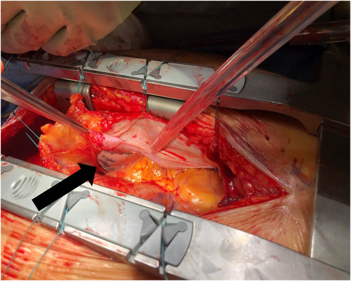
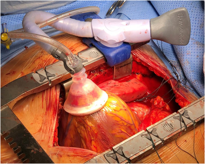
Pericardial rupture with cardiac herniation is a rare traumatic injury with an estimated incidence of 0.37% after blunt trauma. Most commonly occurring after high-speed impact, such as in motor vehicle or motorcycle collisions, pericardial rupture is associated with a high mortality rate. Radiologic diagnosis can be challenging; cross-sectional imaging findings can be suggestive of pericardial rupture but are often non-specific, and echocardiography windows are often obscured. Definitive diagnosis is generally made intra-operatively. Treatment involves reduction of the heart into normal anatomic position with repair of the pericardium, either primarily or with a patch. Fewer than 60 cases of pericardial rupture from blunt trauma have been reported in the literature. We describe a 65 year old poly-trauma patient who sustained pericardial rupture with subsequent cardiac herniation with cardiovascular collapse, and we discuss the considerations and complexities of his successful repair.
Keywords: Cardiac herniation; Cardiac subluxation; Cardiac trauma; Pericardial rupture.
© 2020 The Author(s).
Conflict of interest statement
The views expressed in this manuscript are solely of the authors and do not reflect the views of the United States Air Force or the Department of Defense.
Figures


References
-
- Fulda G., Rodriguez A., Turney S.Z., Cowley R.A. Blunt traumatic pericardial rupture. A ten-year experience 1979 to 1989. J. Cardiovasc. Surg. 1990;31:525–530. - PubMed
