. 2020 May 8;5(3):667-679.
doi: 10.1016/j.bioactmat.2020.04.018. eCollection 2020 Sep.

Gene-activated dermal equivalents to accelerate healing of diabetic chronic wounds by regulating inflammation and promoting angiogenesis

Affiliations

Gene-activated dermal equivalents to accelerate healing of diabetic chronic wounds by regulating inflammation and promoting angiogenesis

Dong Lou et al. Bioact Mater. .

Abstract

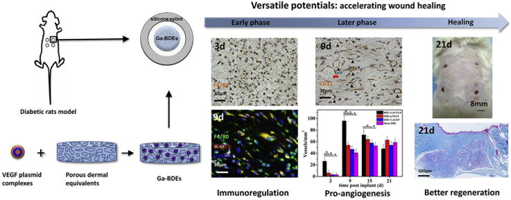
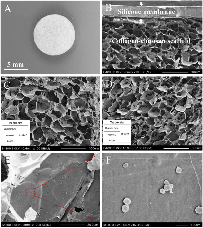
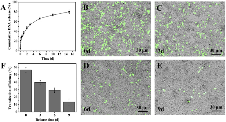
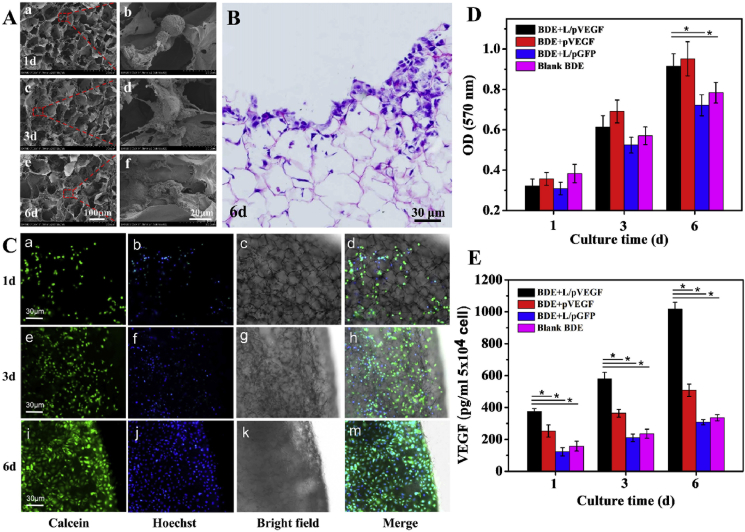
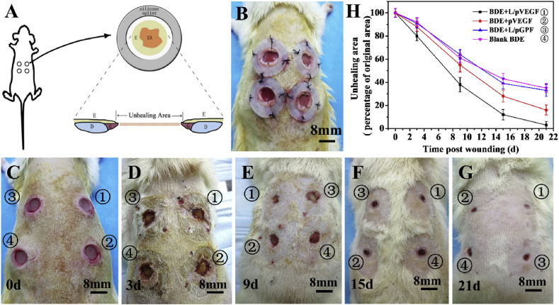
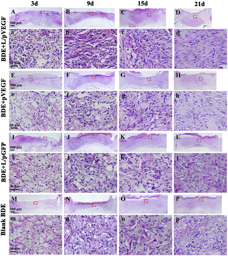
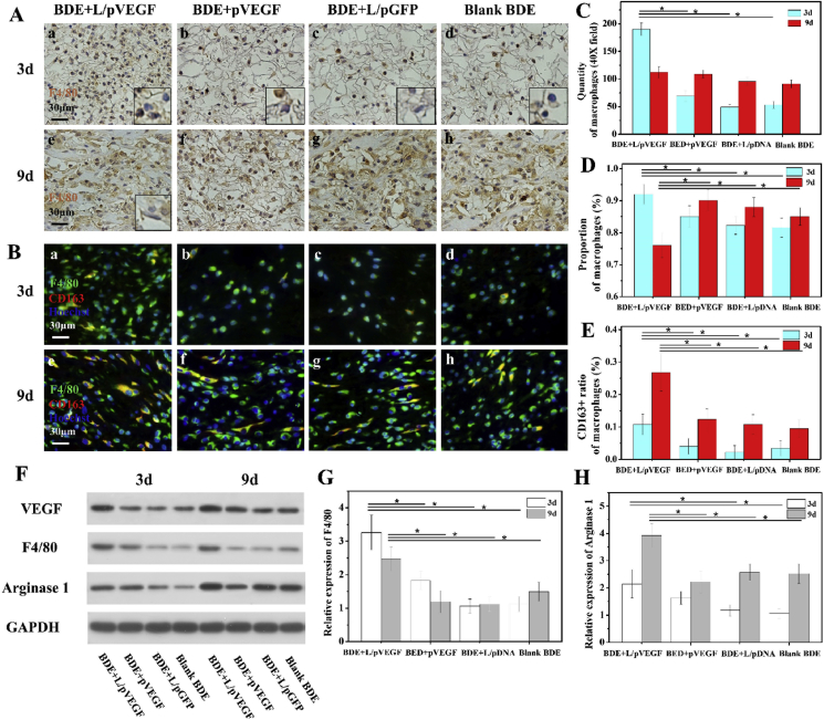
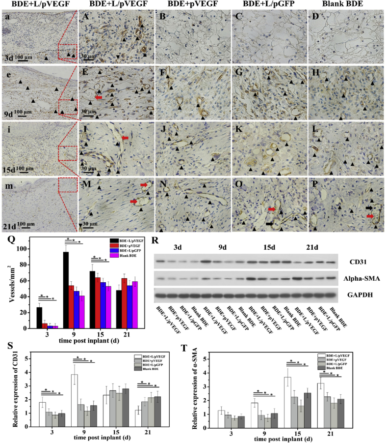
Diabetic chronic wound, characterized by prolonged inflammation and impaired angiogenesis, has become one of the most serious challenges in clinic and pose a significant healthcare burden worldwide. Although a great variety of wound dressings have been developed, few of encouraged achievements were obtained so far. In this study, the gene-activated strategy was applied to enhance sustained expression of vascular endothelial growth factor (VEGF) and achieve better healing outcomes by regulating inflammation and promoting angiogenesis. The gene-activated bilayer dermal equivalents (Ga-BDEs), which has good biocompatibility, were fabricated by loading the nano-sized complexes of Lipofectamine 2000/plasmid DNA-encoding VEGF into a collagen-chitosan scaffold/silicone membrane bilayer dermal equivalent. The DNA complexes were released in a sustained manner and showed the effective transfection capacities to up-regulate the expression of VEGF in vitro. To overcome cutaneous contraction of rodents and mimic the wound healing mechanisms of the human, a reformative rat model of full-thickness diabetic chronic wound was adopted. Under the treatment of Ga-BDEs, speeding wound healing was observed, which is accompanied by the accelerated infiltration and phenotype shift of macrophages and enhanced angiogenesis in early and late healing phases, respectively. These proved that Ga-BDEs possess the functions of immunomodulation and pro-angiogenesis simultaneously. Subsequently, the better regeneration outcomes, including deposition of oriented collagen and fast reepithelialization, were achieved. All these results indicated that, being different from traditional pro-angiogenic concept, the up-regulated expression of VEGF by Ga-BDEs in a sustained manner shows versatile potentials for promoting the healing of diabetic chronic wounds.

Keywords: Angiogenesis; Diabetic chronic wounds; Gene-activated dermal equivalent; Inflammation; Vascular endothelial growth factor.


Conflict of interest statement

The authors declare that they have no known competing financial interests or personal relationships that could have appeared to influence the work reported in this paper.

Figures

Image 1
Graphical abstract
Fig. 1
Fig. 1
Gross view and microstructure of Ga-BDEs. (A) Gross view and (B) SEM image of the gene-activated bilayer dermal equivalent (Ga-BDE). (C) is the SEM image of blank collagen/chitosan scaffold. (D) and (E) are the SEM images of lipofactamine 2000/plasmid complex-loaded collagen/chitosan scaffold with different magnifications. (F) is the enlarged view of the red rectangle area of (E) to show the adhered plasmid complexes.
Fig. 2
Fig. 2
Release pattern and transfection efficiency of released plasmid complexes. (A) Cumulative release of plasmid complexes from BDEs. (B–E) are the fluorescence and bright-field merged images, showing the HEK293 cells transfected by the lipofactamine 2000/pDNA-eGFP complexes released from BDEs at 0, 3, 6 and 9 d, respectively. (F) The quantitative vitro transfection efficiencies, correspondingly.
Fig. 3
Fig. 3
Biocompatibility evaluation and up-regulated VEGF expression. (A) SEM images of NIH 3T3 cells cultured on Ga-BDEs for 1 (a, b), 3 (c, d) and 6 days (e, f). (b), (d) and (f) are the red rectangle-labeled areas in (a), (c) and (e) with higher magnifications, respectively. (B) H&E staining of the Ga-BDE after seeding NIH 3T3 cells for 6 days. (C) The fluorescence, bright-field, and the corresponding merge images of Ga-BDEs after seeding NIH 3T3 cells for 1 (a–d), 3 (e–h), and 6 days (i–m). NIH 3T3 cells were stained by Calcein and Hoechst, respectively. (D) The viabilities of NIH 3T3 cells being cultured in different BDEs for 1, 3, and 6 days, respectively (n = 3). (E) In vitro VEGF expression of HUVECs after being cultured in different BDEs for 1, 3 and 6 days (n = 3). * denotes statistically significant difference at p < 0.05.
Fig. 4
Fig. 4
Reformative animal model and wound healing evaluation. Schematic illustration (A) and representative image (B) of reformative full-thickness incisional model. After creating full-thickness incisional wound, a donut-shaped silicone splint to prevent contraction, was placed and fixed by sutures and tissue glue. The unhealing areas (UA) between bilateral epithelial ingrowth were analyzed using Image J software. E: epithelium, G: granulation tissue, D: dermis. (C–G) are the typical macroscopic observations of the wounds treated by BDEs at different time points. (H) The percentages of UA of the wounds treated by BDEs at different time points. Three wounds at each time point were analyzed to obtain the averaged unhealing ratio.
Fig. 5
Fig. 5
Histological analysis of wound sections treated by different BDEs. H&E stainings of the whole wounds treated by BDEs loaded with L/pVEGF (A–D), pVEGF (E–H) and L/pGFP (I–L), and blank BDE (M-P) for different days, respectively. (a–d), (e–h), (i–l), and (m–p) are the H&E images with higher magnifications, correspondingly.
Fig. 6
Fig. 6
Macrophages analysis and western blotting test. (A) Immunohistochemical staining of F4/80 of the wounds treated by different BDEs for 3 (A, a-d) and 9 days (A, e-h). The inserts of (A, a-d) are the corresponding detail with higher magnification to show F4/80 negative neutrophils-like cells with lobular nucleus. The insert of (A, e) shows the F4/80 negative fibroblasts-like cells with fusiform nucleus. (B) Triple immunofluorescence of wounds sections treated by different BDEs for 3 (B, a-d) and 9d (B, e-h). Statistics of the quantities (C) and proportions (D) of macrophages, and the proportions of CD163+ macrophages (E) in different groups at day 3 and 9 (n ≥ 3). (F) Western blotting (WB) of VEGF, F4/80, and Arginase 1 expressions of the wounds treated by different BDEs at different time points. (G, H) Densitometry analyses of the Western blots of F4/80 and Arginase 1 (n = 3). * denotes statistically significant difference at p < 0.05.
Fig. 7
Fig. 7
Pro-angiogenic effect of different BDEs. Immunohistological stainings of CD 31 of the wounds treated by different BDEs for 3 (A–D), 9 (E–H), 15 (I–L), and 21 days (M-P). Red and black arrows show mature vessels with erythrocyte flow and the inflammatory cell being recruited from circulation, respectively. (a, e, i, m) are the overview images with lower magnification of (A, E, I, M) to show the newly-formed blood vessels in L/pVEGF group. Black triangle showed CD 31+ blood vessels. (Q)The statistic numbers of the newly-formed blood vessels (CD31+ positive stainings) per mm2 (n ≥ 6). (R) WB of CD31 and α-SMA expressions of the wounds treated by different BDEs at different time points. (S, T) Densitometry analyses of the Western blots of CD31 and α-SMA (n = 3). * denotes statistically significant difference at p < 0.05.
Fig. 8
Fig. 8
Assessment of tissue remodeling by Masson's staining. Masson's stainings of the whole wounds treated by different BDEs in later healing phase at 15 d (A–D) and 21 d (E–H). (a–d) and (e–h) are the magnified images of the red rectangle-labeled areas in (A–D) and (E–H), correspondingly. Black arrows show epithelial gaps.

References

    1. Ogurtsova K., Da R.F.J., Huang Y., Linnenkamp U., Guariguata L., Cho N.H., Cavan D., Shaw J.E., Makaroff L.E. IDF Diabetes Atlas: global estimates for the prevalence of diabetes for 2015 and 2040. Diabetes Res. Clin. Pract. 2017;128:40–50. - PubMed
    1. Baltzis D., Eleftheriadou I., Veves A. Pathogenesis and treatment of impaired wound healing in diabetes mellitus: new insights. Adv. Ther. 2014;31:817–836. - PubMed
    1. Boulton A.J. The pathway to foot ulceration in diabetes. Med Clin North Am. 2013;97:775–790. - PubMed
    1. Mulder G.D., Patt L.M., Sanders L., Rosenstock J., Altman M.I., Hanley M.E., Duncan G.W. Enhanced healing of ulcers in patients with diabetes by topical treatment with glycyl-l-histidyl-l-lysine copper. Wound Repair Regen. 1994;2:259–269. - PubMed
    1. Falanga V. Wound healing and its impairment in the diabetic foot. Lancet. 2005;366:1736–1743. - PubMed

LinkOut - more resources