. 2020 Aug 1;77(8):937-946.
doi: 10.1001/jamaneurol.2020.1162.

Association Between Tumor Necrosis Factor Inhibitor Exposure and Inflammatory Central Nervous System Events

Affiliations

Association Between Tumor Necrosis Factor Inhibitor Exposure and Inflammatory Central Nervous System Events

Amy Kunchok et al. JAMA Neurol. .

Abstract

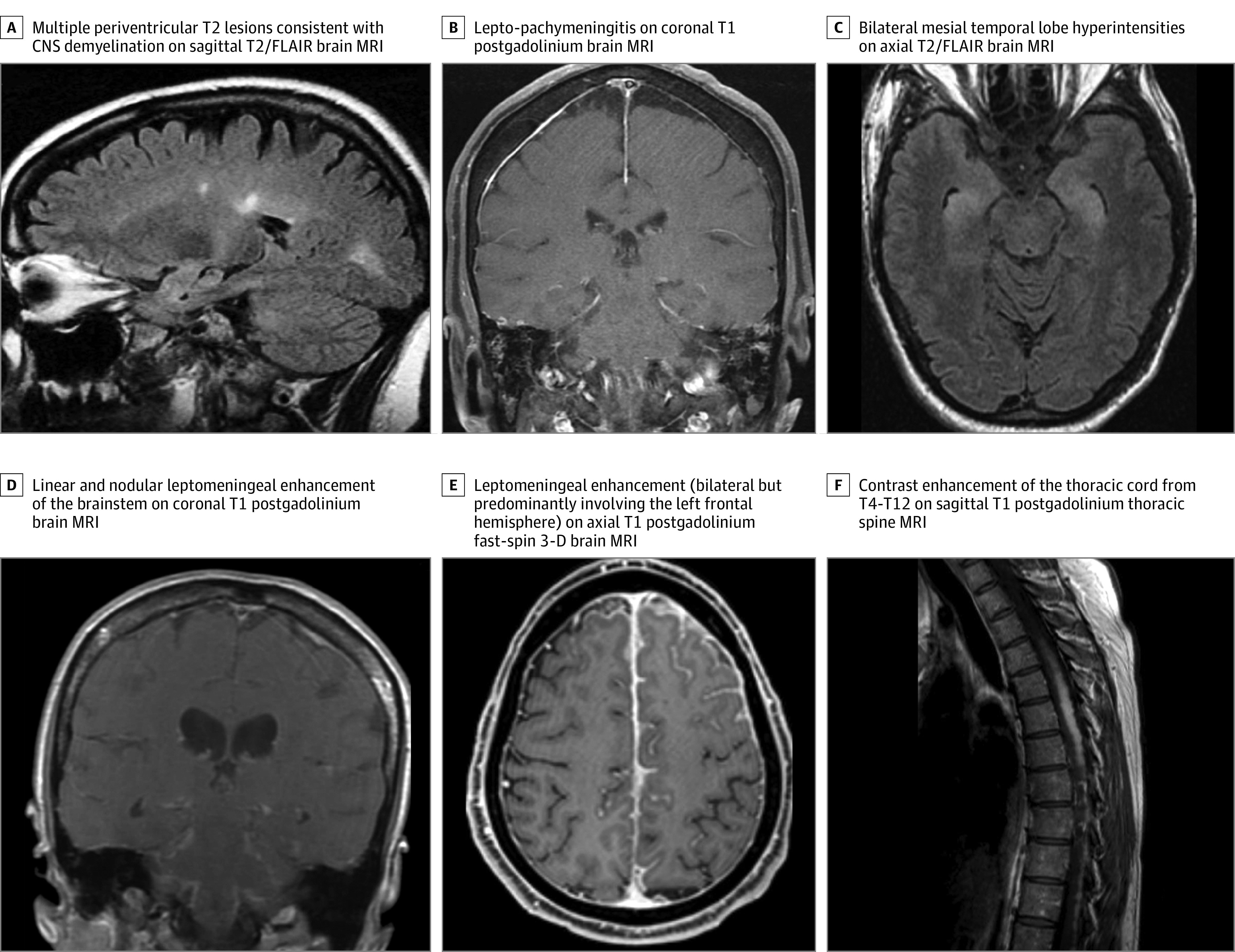
Importance: Tumor necrosis factor (TNF) inhibitors are common therapies for certain autoimmune diseases, such as rheumatoid arthritis. An association between TNF inhibitor exposure and inflammatory central nervous system (CNS) events has been postulated but is poorly understood.

Objective: To evaluate whether TNF inhibitor exposure is associated with inflammatory demyelinating and nondemyelinating CNS events in patients with an indication for TNF inhibitor use and to describe the spectrum of those CNS events.

Design, setting, and participants: A nested case-control study was conducted using the medical records of patients with autoimmune diseases treated at 3 Mayo Clinic locations (Rochester, Minnesota; Scottsdale, Arizona; and Jacksonville, Florida) between January 1, 2003, and February 20, 2019. Patients were included if their records reported International Statistical Classification of Diseases and Related Health Problems, Tenth Revision, diagnostic codes for US Food and Drug Administration-approved autoimmune disease indication for TNF inhibitor use (ie, rheumatoid arthritis, ankylosing spondylitis, psoriasis and psoriatic arthritis, Crohn disease, and ulcerative colitis) and diagnostic codes for inflammatory CNS events of interest. Patients were matched 1:1 with control participants by year of birth, type of autoimmune disease, and sex.

Exposures: TNF inhibitor exposure data were derived from the medical records along with type of TNF inhibitor, cumulative duration of exposure, and time of exposure.

Main outcomes and measures: The main outcome was either inflammatory demyelinating (multiple sclerosis and other diseases such as optic neuritis) or nondemyelinating (meningitis, meningoencephalitis, encephalitis, neurosarcoidosis, and CNS vasculitis) CNS event. Association with TNF inhibitor was evaluated with conditional logistic regression and adjusted for disease duration to determine the odds ratios (ORs) and 95% CIs. Secondary analyses included stratification of outcome by inflammatory demyelinating and nondemyelinating CNS events and by autoimmune disease (rheumatoid arthritis and non-rheumatoid arthritis).

Results: A total of 212 individuals were included: 106 patients with inflammatory CNS events and 106 control participants without such events. Of this total, 136 were female (64%); the median (interquartile range) age at disease onset for patients was 52 (43-62) years. Exposure to TNF inhibitors occurred in 64 patients (60%) and 42 control participants (40%) and was associated with an increased risk of any inflammatory CNS event (adjusted OR, 3.01; 95% CI, 1.55-5.82; P = .001). These results were similar when the outcome was stratified by demyelinating and nondemyelinating CNS events. Secondary analyses found the association was predominantly observed in patients with rheumatoid arthritis (adjusted OR, 4.82; 95% CI, 1.62-14.36; P = .005).

Conclusions and relevance: This study found that exposure to TNF inhibitors in patients with autoimmune diseases appeared to be associated with increased risk for inflammatory CNS events. Whether this association represents de novo or exacerbated inflammatory pathways requires further research.


Conflict of interest statement

Conflict of Interest Disclosures: Dr Kunchok reported receiving research funding from Biogen outside the submitted work. Dr Kantarci reported receiving grants from Biogen outside the submitted work. Dr Davis reported receiving grants from Pfizer and Genentech as well as serving on advisory boards for both Abbvie and Sanofi-Genzyme. Dr Pittock reported receiving grants and personal fees from Alexion and Viela Bio, personal fees from Union Chimique Belge and Astellas, and grants from Griffols and Autoimmune Encephalitis Alliance outside the submitted work. Dr Weinshenker reported receiving personal fees from Viela Bio, Alexion, Chugai/Roche, Mitsubishi Tanabe, Novartis, Caladrius, Brainstorm Therapeutics, and Roivant outside the submitted work, as well as holding a patent to NMO-IgG for diagnosis of neuromyelitis optica that has been issued and licensed and with paid royalties. Dr McKeon reported receiving grants from Euroimmun and Medimmune outside the submitted work; being a member of the editorial board of Neurology: Neuroimmunology and Neuroinflammation; holding pending patent applications for Septin 5, GFAP, PDE10A, Kelch-11, and MAP1B IgGs as markers of neurological autoimmunity and paraneoplastic disorders; and receiving research support from Euroimmun, Grifols, and Alexion. No other disclosures were reported.

Figures

Figure.
Figure.. Magnetic Resonance Imaging (MRI) Features of Some Patients
A, A patient with Crohn disease for 3 years who was treated with adalimumab for 10 years presented with a thoracic sensory band and lower-limb paresthesia. The patient had a cervical spine MRI that revealed multiple short-segment T2 hyperintense lesions with contrast enhancement. The diagnosis was central nervous system (CNS) demyelination. B, A patient with a history of rheumatoid arthritis who was treated with adalimumab for 16 years presented with headaches and a generalized tonic-clonic seizure; an infectious and malignant neoplasm evaluation of cerebrospinal fluid (CSF) had a negative result. Dural biopsy results revealed necrotizing meningitis, and were negative for special stains for microorganisms, including mycobacteria and fungi. The diagnosis was idiopathic lepto-pachymeningitis. C, A patient with ankylosing spondylitis and type 1 diabetes was treated with several tumor necrosis factor (TNF) inhibitors for a cumulative total of 2 years (infliximab twice, etanercept, and adalimumab). The patient had ceased using TNF inhibitor 10 months before presenting with acute confusion. The CSF analysis and positron emission tomography (PET) scan had a negative result for neural antibodies and malignant neoplasm. Serum autoimmune neural antibodies had a positive result (serum acetylcholine receptor modulating antibody, 34% loss; GAD65 antibody, 0.35 nmol/L; striational antibody, 1:240). The diagnosis was autoimmune encephalitis. The patient had a clinically robust response to intravenous methylprednisolone acetate. D, A patient with Crohn disease for 10 years was treated with adalimumab for 6 months, then infliximab for 6 months before presenting with new-onset headaches, hearing loss, paresthesias, left facial droop, numbness and diplopia. In addition to a brain MRI scan showing leptomeningeal enhancement of the brainstem, an MRI of the cervical and thoracic spine also revealed patchy leptomeningeal enhancement of the spinal cord. Results of an infectious and neoplastic evaluation of the CSF were negative. A PET scan identified increased fluorodeoxyglucose (FDG) uptake in the mediastinum, and a biopsy of subcarinal lymph node confirmed granulomatous disease. Special stains for microorganisms, including mycobacteria and fungi, had a negative result. The diagnosis was neurosarcoidosis. E, A patient with rheumatoid arthritis for 8 years who was treated with etanercept and methotrexate sodium presented with new-onset headaches and paresthesia of the right face and limbs. A right frontal biopsy specimen demonstrated necrotizing granulomatous inflammation that extensively involved the leptomeninges. Special stains for microorganisms, including mycobacteria and fungi, had a negative result. The diagnosis was neurosarcoidosis. F, A patient with a history of Crohn disease who was treated with infliximab for 3 years presented with myelopathic symptoms. A PET scan showed moderately intense FDG activity in the thoracic spinal cord. A thoracic cord biopsy demonstrated necrotizing granulomatous inflammation. Special stains for microorganisms, including mycobacteria and fungi, had a negative result. The diagnosis was neurosarcoidosis. FLAIR indicates fluid-attenuated inversion recovery sequence.

Comment in

References

    1. Singh S, Garg SK, Pardi DS, Wang Z, Murad MH, Loftus EV Jr. Comparative efficacy of pharmacologic interventions in preventing relapse of Crohn’s disease after surgery: a systematic review and network meta-analysis. Gastroenterology. 2015;148(1):64-76.e2. doi:10.1053/j.gastro.2014.09.031 - DOI - PMC - PubMed
    1. Elliott MJ, Maini RN, Feldmann M, et al. . Randomised double-blind comparison of chimeric monoclonal antibody to tumour necrosis factor alpha (cA2) versus placebo in rheumatoid arthritis. Lancet. 1994;344(8930):1105-1110. doi:10.1016/S0140-6736(94)90628-9 - DOI - PubMed
    1. Slevin SM, Egan LJ. New insights into the mechanisms of action of anti-tumor necrosis factor-α monoclonal antibodies in inflammatory bowel disease. Inflamm Bowel Dis. 2015;21(12):2909-2920. doi:10.1097/MIB.0000000000000533 - DOI - PubMed
    1. van Oosten BW, Barkhof F, Truyen L, et al. . Increased MRI activity and immune activation in two multiple sclerosis patients treated with the monoclonal anti-tumor necrosis factor antibody cA2. Neurology. 1996;47(6):1531-1534. doi:10.1212/WNL.47.6.1531 - DOI - PubMed
    1. The Lenercept Multiple Sclerosis Study Group and The University of British Columbia MS/MRI Analysis Group TNF neutralization in MS: results of a randomized, placebo-controlled multicenter study. Neurology. 1999;53(3):457-465. doi:10.1212/WNL.53.3.457 - DOI - PubMed

Publication types

MeSH terms

Substances