. 2020 Jun 19;15(6):1581-1594.
doi: 10.1021/acschembio.0c00184. Epub 2020 May 18.

Structure-Guided Optimization of Inhibitors of Acetyltransferase Eis from Mycobacterium tuberculosis

Affiliations

Structure-Guided Optimization of Inhibitors of Acetyltransferase Eis from Mycobacterium tuberculosis

Ankita Punetha et al. ACS Chem Biol. .

Abstract

The enhanced intracellular survival (Eis) protein of Mycobacterium tuberculosis (Mtb) is a versatile acetyltransferase that multiacetylates aminoglycoside antibiotics abolishing their binding to the bacterial ribosome. When overexpressed as a result of promoter mutations, Eis causes drug resistance. In an attempt to overcome the Eis-mediated kanamycin resistance of Mtb, we designed and optimized structurally unique thieno[2,3-d]pyrimidine Eis inhibitors toward effective kanamycin adjuvant combination therapy. We obtained 12 crystal structures of enzyme-inhibitor complexes, which guided our rational structure-based design of 72 thieno[2,3-d]pyrimidine analogues divided into three families. We evaluated the potency of these inhibitors in vitro as well as their ability to restore the activity of kanamycin in a resistant strain of Mtb, in which Eis was upregulated. Furthermore, we evaluated the metabolic stability of 11 compounds in vitro. This study showcases how structural information can guide Eis inhibitor design.


Conflict of interest statement

The authors declare no competing financial interest.

Figures

Figure 1.
Figure 1.
Cone diagram showing the evaluation and triage of ∼23 000 structurally diverse compounds.
Figure 2.
Figure 2.
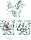
Crystal structures of Eis (one monomer is shown) in complex with inhibitors. (A) Crystal structure of Eis−2i complex. Eis is shown in cyan and 2i as orange sticks. (B) Zoomed-in view of the Eis−2i interface. The polder omit map for the inhibitor is contoured at 4σ and is shown by the brown mesh. The amino acid residues interacting with compound 2i are shown by dark turquoise sticks. The nitrogen atoms are in light blue, oxygen atoms in red, sulfur atoms in yellow, and fluorine atoms in cyan. The C-terminus is labeled “C-ter”. (C) Previously reported crystal structure of Eis−39b complex (PDB ID 6B3T). A water molecule in the active site is shown as a red sphere. The chemical structures of the bound inhibitors are shown on the bottom of panels B and C. Note: This color scheme is preserved in all the other figures depicting crystal structures.
Figure 3.
Figure 3.
Chemical structures of all compounds synthesized and investigated in this study. The three rings of the tricyclic inhibitors in the library are labeled as A, B, and C for simplicity. The library is divided into three families (I−III) based on the size of ring A. Further subdivision of the families is based on R1, R2, and R3 substituents.
Figure 4.
Figure 4.
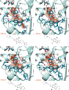
Zoomed-in views of the Eis−inhibitor interfaces from crystal structures of Eis−1g and Eis−2g complexes, and the inhibitor structures: (A) Eis−1g interface and (B) Eis−2g interface.
Figure 5.
Figure 5.
Zoomed-in views of the Eis−inhibitor interfaces from crystal structures of Eis−1h and Eis−8h complexes and the inhibitor structures. (A) Eis−1h interface and (B) Eis−2h interface.
Figure 6.
Figure 6.
Zoomed-in views of the Eis−inhibitor interfaces from crystal structures of Eis−2e and Eis−4e complexes and the inhibitor structures: (A) Eis−2e interface and (B) Eis−4e interface.
Figure 7.
Figure 7.
Zoomed-in views of the Eis−inhibitor interfaces from crystal structures of Eis−7c, Eis−1c, Eis−4c, and Eis−8c complexes and the inhibitor structures: (A) Eis−7c interface, (B) Eis−1c interface, (C) Eis−4c interface, and (D) Eis−8c interface.
Figure 8.
Figure 8.
Zoomed-in view of the Eis−inhibitor interface from crystal structure of Eis−5h complex and the structure of 5h.
Figure 9.
Figure 9.
Analysis of inhibition kinetics for inhibitors 4c, 7c, and 4g. Dose−response curves are shown in panels A, D, and G; Michaelis−Menten analysis is in panels B, E, and H; and the Lineweaver−Burk representation of this analysis is in panels C, F, and I. These data indicate that the molecules are competitive with KAN. These data are used in obtaining Ki by global nonlinear regression (Table 2).
Figure 10.
Figure 10.
Evaluation of cytotoxicity for compounds 2e (red), 2i (yellow), 7c (purple), and KAN (gray) against three cell lines: (A) A549, (B) HEK-293, and (C) J774A.1. Controls include treatment with Triton-X (TX, 1% v/v, positive control) and 0.5% DMSO (negative control). It is important to note that testing xenobiotics at sub-IC50 concentrations can result in increase in cell growth, resulting in >100% cell survival in the treatment groups. In instances where >100% cell survival was observed, we displayed the data as 100% cell survival. Note: The raw data are presented in Figure S230.
Scheme 1.
Scheme 1.
Synthetic Scheme for the Preparation of (A) Compounds 1−8(a−i) and (B) Intermediates 41−44

References

    1. Rice LB (2008) Federal funding for the study of antimicrobial resistance in nosocomial pathogens: no ESKAPE. J. Infect. Dis 197, 1079–1081. - PubMed
    1. Global Tuberculosis Report 2019, World Health Organization, Geneva, Switzerland, 2019, licence CCBY-NC-SA3.01GO.
    1. Zaunbrecher MA, Sikes RD Jr., Metchock B, Shinnick TM, and Posey JE (2009) Overexpression of the chromosomally encoded aminoglycoside acetyltransferase eis confers kanamycin resistance in Mycobacterium tuberculosis. Proc. Natl. Acad. Sci. U. S. A 106, 20004–20009. - PMC - PubMed
    1. Campbell PJ, Morlock GP, Sikes RD, Dalton TL, Metchock B, Starks AM, Hooks DP, Cowan LS, Plikaytis BB, and Posey JE (2011) Molecular detection of mutations associated with first- and second-line drug resistance compared with conventional drug susceptibility testing of Mycobacterium tuberculosis. Antimicrob. Agents Chemother 55, 2032–2041. - PMC - PubMed
    1. Reeves AZ, Campbell PJ, Sultana R, Malik S, Murray M, Plikaytis BB, Shinnick TM, and Posey JE (2013) Aminoglycoside cross-resistance in Mycobacterium tuberculosis due to mutations in the 5′ untranslated region of whiB7. Antimicrob. Agents Chemother 57, 1857–1865. - PMC - PubMed

Publication types

MeSH terms