. 2020 May 18;15(5):e0232849.
doi: 10.1371/journal.pone.0232849. eCollection 2020.

Sequence-structure-function relationships in class I MHC: A local frustration perspective

Affiliations

Sequence-structure-function relationships in class I MHC: A local frustration perspective

Onur Serçinoğlu et al. PLoS One. .

Abstract

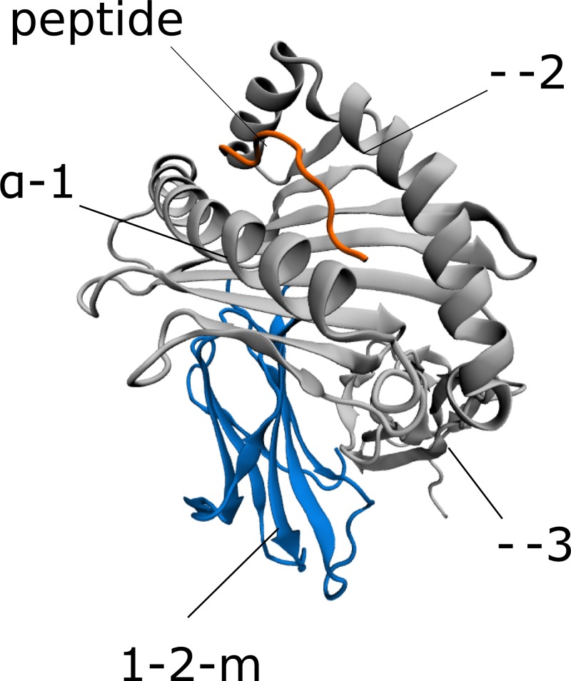
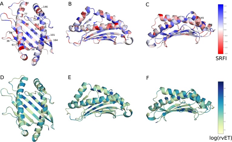
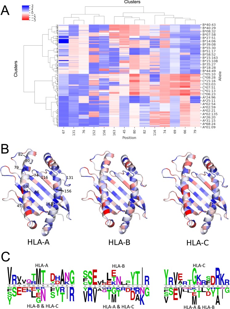
Class I Major Histocompatibility Complex (MHC) binds short antigenic peptides with the help of Peptide Loading Complex (PLC), and presents them to T-cell Receptors (TCRs) of cytotoxic T-cells and Killer-cell Immunglobulin-like Receptors (KIRs) of Natural Killer (NK) cells. With more than 10000 alleles, human MHC (Human Leukocyte Antigen, HLA) is the most polymorphic protein in humans. This allelic diversity provides a wide coverage of peptide sequence space, yet does not affect the three-dimensional structure of the complex. Moreover, TCRs mostly interact with HLA in a common diagonal binding mode, and KIR-HLA interaction is allele-dependent. With the aim of establishing a framework for understanding the relationships between polymorphism (sequence), structure (conserved fold) and function (protein interactions) of the human MHC, we performed here a local frustration analysis on pMHC homology models covering 1436 HLA I alleles. An analysis of local frustration profiles indicated that (1) variations in MHC fold are unlikely due to minimally-frustrated and relatively conserved residues within the HLA peptide-binding groove, (2) high frustration patches on HLA helices are either involved in or near interaction sites of MHC with the TCR, KIR, or tapasin of the PLC, and (3) peptide ligands mainly stabilize the F-pocket of HLA binding groove.


Conflict of interest statement

The authors have declared that no competing interests exist.

Figures

Fig 1
Fig 1. The three-dimensional structure of the pMHC.
Fig 2
Fig 2. Sequence conservation/variation and evolutionary importance of HLA I peptide-binding groove positions.
(A) Sequence logo of the HLA I peptide-binding groove (residues 1–180). Polar, neutral, basic, acidic and hydrophobic amino acids are colored green, purple, blue, red, and black, respectively. (B) real-value Evolutionary Trace (rvET) scores of binding groove positions. Low rvET scores indicate high evolutionary importance, and vice versa.
Fig 3
Fig 3. Sequence conservation/variation and evolutionary importance of HLA I α-3 domain positions.
(A) Sequence logo of the HLA I α-3 domain (residues 181–261). Polar, neutral, basic, acidic and hydrophobic amino acids are colored green, purple, blue, red, and black, respectively. (B) real-value Evolutionary Trace (rvET) scores of α-3 domain positions. Low rvET scores indicate high evolutionary importance, and vice versa.
Fig 4
Fig 4. Position-specific Single Residue Frustration Index (SRFI) and rvET scores mapped onto the HLA I binding groove.
(A, B, C) SRFI mapped onto binding groove positions. (D, E, F) log(rvET) mapped onto binding groove positions. Selected positions are also shown on the structure.
Fig 5
Fig 5. Box-plots of position-specific SRFI values for the HLA I peptide-binding groove.
(A) SRFI box-plots of selected minimally and highly frustrated residues within the HLA peptide binding groove (B) SRFI box-plots of positions with neutral frustration. (C) SRFI box-plots of peptide-binding pocket positions. Coloring according to log(rvET) values.
Fig 6
Fig 6. Position-specific Single Residue Frustration Index (SRFI) and rvET scores mapped onto the HLA I α-3 domain.
(A) SRFI mapped onto α-3 domain positions. (B) log(rvET) mapped onto α-3 domain positions. Selected positions are also shown on the structure.
Fig 7
Fig 7. Box-plots of position-specific SRFI values for the HLA I α-3 domain.
SRFI box-plots of minimally- and highly-frustrated residues within the HLA α-3 domain. Coloring according to log(rvET) values.
Fig 8
Fig 8. Minimally-frustrated and conserved positions within the HLA structure.
Residues are drawn in van der Waals spheres representation. Coloring according to SRFI value. The structure of HLA-B*53:01 (1A1M) was used for demonstration purposes. Selected β2m residues are also shown in spheres to highlight interaction with HLA.
Fig 9
Fig 9. Clustering of allele-specific SRFI profiles based on 14 binding groove positions.
(A) SRFI cluster heatmap. Clustering was performed for all 1436 alleles included, yet not all of these alleles are indicated on axis label for clarity. (B) SRFI of three identified clusters corresponding to three HLA gene loci mapped onto the binding groove. Coloring as in Fig 5. (C) Sequence logos of clustering positions to highlight amino acid differences between allele in different clusters. The logos were generated using Two Sample Logo server [86].
Fig 10
Fig 10. Box-plots of SRFI change upon peptide binding.
Positions are ordered from left to right according to decreasing change in median SRFI values. Note that the plot only includes top 10 positions showing the highest increase in SRFI levels upon peptide binding.
Fig 11
Fig 11. Clustering of allele-specific SRFI profiles based on.
(A) peptide-binding pocket and (B) TAPBPR interface residues of 30 TAPBPR binders and non-binders as reported by Ilca et al. [101]. Position 116 was also included in clustering shown in B. TAPBPR interface residues were based on a previously reported structure of a TAPBPR-MHC I complex. Top TAPBPR binders are highlighted with dashed-line rectangles.

References

    1. Liberles DA, Teichmann SA, Bahar I, Bastolla U, Bloom J, Bornberg-Bauer E, et al. The interface of protein structure, protein biophysics, and molecular evolution Protein Science. Wiley-Blackwell; 2012. pp. 769–785. 10.1002/pro.2071 - DOI - PMC - PubMed
    1. Bastolla U, Dehouck Y, Echave J. What evolution tells us about protein physics, and protein physics tells us about evolution Current Opinion in Structural Biology. Elsevier Current Trends; 2017. pp. 59–66. 10.1016/j.sbi.2016.10.020 - DOI - PubMed
    1. Bahar I, Jernigan RL, Dill KA. Protein actions: principles and modeling. 2017. Available: https://www.crcpress.com/Protein-Actions-Principles-and-Modeling/Bahar-J...
    1. Kimura M, Ohta T. On some principles governing molecular evolution. Proc Natl Acad Sci U S A. 1974;71: 2848–52. Available: http://www.ncbi.nlm.nih.gov/pubmed/4527913 10.1073/pnas.71.7.2848 - DOI - PMC - PubMed
    1. Parra RG, Espada R, Verstraete N, Ferreiro DU. Structural and Energetic Characterization of the Ankyrin Repeat Protein Family. Orengo CA, editor. PLoS Comput Biol. 2015;11: e1004659 10.1371/journal.pcbi.1004659 - DOI - PMC - PubMed

Publication types