. 2020 Jun 19;48(11):5891-5906.
doi: 10.1093/nar/gkaa344.

Che-1/AATF binds to RNA polymerase I machinery and sustains ribosomal RNA gene transcription

Affiliations

Che-1/AATF binds to RNA polymerase I machinery and sustains ribosomal RNA gene transcription

Cristina Sorino et al. Nucleic Acids Res. .

Abstract

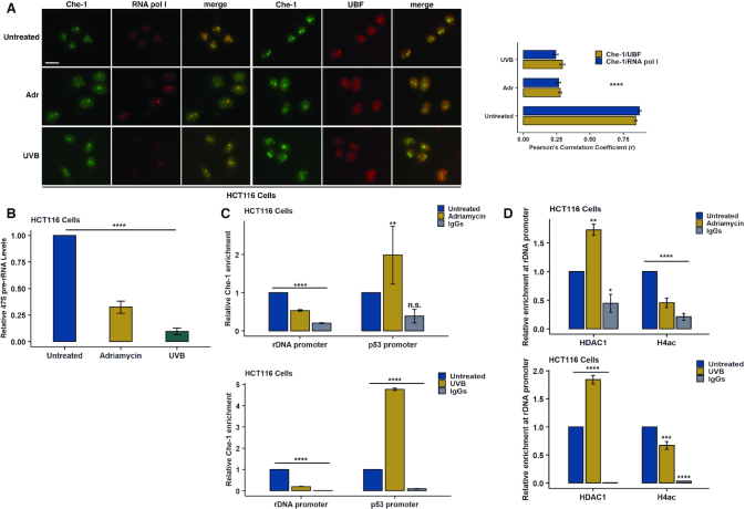
Originally identified as an RNA polymerase II interactor, Che-1/AATF (Che-1) has now been recognized as a multifunctional protein involved in cell-cycle regulation and cancer progression, as well as apoptosis inhibition and response to stress. This protein displays a peculiar nucleolar localization and it has recently been implicated in pre-rRNA processing and ribosome biogenesis. Here, we report the identification of a novel function of Che-1 in the regulation of ribosomal RNA (rRNA) synthesis, in both cancer and normal cells. We demonstrate that Che-1 interacts with RNA polymerase I and nucleolar upstream binding factor (UBF) and promotes RNA polymerase I-dependent transcription. Furthermore, this protein binds to the rRNA gene (rDNA) promoter and modulates its epigenetic state by contrasting the recruitment of HDAC1. Che-1 downregulation affects RNA polymerase I and UBF recruitment on rDNA and leads to reducing rDNA promoter activity and 47S pre-rRNA production. Interestingly, Che-1 depletion induces abnormal nucleolar morphology associated with re-distribution of nucleolar proteins. Finally, we show that upon DNA damage Che-1 re-localizes from rDNA to TP53 gene promoter to induce cell-cycle arrest. This previously uncharacterized function of Che-1 confirms the important role of this protein in the regulation of ribosome biogenesis, cellular proliferation and response to stress.


Figures

Figure 1.
Figure 1.
Che-1 interacts with RNA pol I transcription machinery and binds to rDNA. (A) Scatter plot of putative RNA pol I interacting proteins. The results of the t-test comparison of protein intensities measured via MS are depicted. On the x-axis is the mean log2 difference in intensities of proteins measured in IP RNA pol I (subunit RPA194) versus IP control; on the y-axis are the corresponding −log10P-values. In black are indicated the known components of the RNA pol I complex identified in the experiment and the protein Che-1 (AATF). For a complete list of all significant interactors, please refer to Supplementary Table S1. (B) Nuclear extracts from HCT116 cells were subjected to immunoprecipitation with either RNA pol I (RPA194 subunit; top) or UBF (bottom) antibodies. Immuno-precipitated complexes were then analysed by WB with the indicated antibodies. Input corresponds to 5% of the nuclear extract used for immunoprecipitation. (C) Representative images showing the interaction between either RNA pol I and Che-1 or UBF and Che-1 revealed by PLA. Negative controls were performed by omitting Che-1 primary antibody, whilst nuclei were stained with Hoechst dye. Scale bar, 10 μM. (D) Nuclear extracts from HCT116 cells were centrifuged on a linear 10–30% sucrose gradient. Equal volumes of the collected fractions were analysed by WB with the indicated antibodies. (E) Left: HCT116 nuclear extracts were immunoprecipitated with either RNA pol I (RPA194 subunit; top) or Che-1 (bottom) antibodies in the presence or absence of RNAse A 20 μg/ml. Immuno-precipitated complexes were then analysed by WB with the indicated antibodies. Input corresponds to 5% of the nuclear extract used for immunoprecipitation. Right: Relative Che-1 binding (top) and UBF binding (bottom) were calculated from three different experiments by densitometry. Che-1 and UBF intensities were normalized to the ones of RNA pol I and Che-1, respectively. (F) Top: Che-1 occupancy at rDNA locus was evaluated by ChIP assays with a specific Che-1 antibody in HCT116 cells. The percentage of precipitated DNA was evaluated by real-time PCR with the indicated primer sets and calculated based on the ChIP input. Bottom: Schematic representation of an individual rDNA repeat. (G) Re-ChIP experiments were performed in HCT116 cells with the indicated antibodies. The percentage of precipitated DNA was evaluated by real-time PCR with a specific primer set for the H42.9 region and calculated based on the ChIP input. Data are presented as percentage of first round of ChIP. Statistical significance is indicated by asterisks as follows: *P< 0.05, **P< 0.01, ***P< 0,005, ****P< 0,001, n.s. = not significant. Please also see Supplementary Figure S1.
Figure 2.
Figure 2.
Che-1 promotes rDNA transcription. (A) Schematic representation of human 47S pre-rRNA. 5’ ETS, 5’ external transcribed region; ITS1, internal transcribed region 1; ITS2, internal transcribed region 2; 3’ ETS, 3’ external transcribed region. (B) Real-time PCR (qRT-PCR) analysis of 47S pre-rRNA levels in the indicated cell lines transfected with siRNA oligonucleotides targeting Che-1 (siChe-1) or a control sequence (siControl). Relative fold changes were determined by the comparative threshold (ΔΔCT) method using β-actin as endogenous normalization control. (C) Luciferase activity of human rDNA promoter (pHrD-IRES-luc) was measured in the indicated cell lines transfected with pHrD-IRES-luc and siChe-1 or siControl oligonucleotides. (D) Left: 5-FUrd labelled RNA was evaluated by immunostaining with anti-BrdU antibody in HCT116 cells transfected with two different siRNA oligonucleotides targeting Che-1 (siChe-1 and siChe-1 b) or a control sequence (siControl). Nuclei were stained with Hoechst dye. Scale bar, 10 μM. Right: bar plot showing the percentage of 5-FUrd positive nuclei (top). Percentage was calculated from 100 randomly selected cells for each group. Representative WB showing the transfection efficiency of the 5-FUrd incorporation assay described above (bottom). The bar plot under the WB shows the average downregulation of Che-1 observed in these experiments (n = 3). (E) qRT-PCR analysis of 47S pre-rRNA levels in the indicated cell lines transfected with either empty vector or Che-1 expressing vector (Che-1 myc). Relative fold changes were determined by the comparative threshold (ΔΔCT) method using β-actin as endogenous normalization control. (F) Luciferase activity of human rDNA promoter was measured in the indicated cell lines transfected with pHrD-IRES-luc along with empty vector or Che-1 expressing vector (Che-1 myc). (G) Luciferase activity of human rDNA promoter was measured in BJ human fibroblasts transfected with siChe-1 or siControl oligonucleotides or with empty vector or Che-1 expressing vector (Che-1 myc). All data are expressed as a percentage of control value (siControl/empty vector) and presented as mean ± SD of three independent experiments. Statistical significance is indicated by asterisks as follows: *P< 0.05, **P< 0.01, ***P< 0.005, ****P< 0.001, n.s. = not significant. Please also see Supplementary Figure S2.
Figure 3.
Figure 3.
Che-1 affects rDNA epigenetic state and modulates UBF and RNA pol I binding to the rDNA promoter. (A) Luciferase activity of human rDNA promoter was measured in 293T cells transfected with pHrD-IRES-luc and either empty vector or HDAC1 expressing vector (HDAC1 myc) alone or in combination with Che-1 expressing vector (Che-1 myc). Data are expressed as a percentage of the control value (empty vector) and presented as mean ± SD of three independent experiments, performed in triplicate (top). WB analysis with the indicated antibodies of representative samples used in the luciferase assay showed above (bottom). (B) Representative WB with the indicated antibodies of total cellular extracts from HCT116 cells transfected with siChe-1 or siControl oligonucleotides and simultaneously subjected to the ChIP analyses described below. The bar plot under the WB shows the average downregulation of Che-1 observed in these experiments (n = 3). (C) HDAC1 occupancy at rDNA promoter was evaluated by ChIP assay with a specific HDAC1 antibody in HCT116 cells depleted or not for Che-1 expression. The amount of precipitated DNA was evaluated by real-time PCR with a specific primer set for the H42.9 region. (D) Epigenetic state of rDNA promoter was evaluated by ChIP assays with the indicated antibodies in HCT116 cells depleted or not for Che-1 expression. The amount of precipitated DNA was evaluated by real-time PCR with a specific primer set for the H42.9 region. RNA pol I (E) and UBF (F) occupancy at rDNA locus was evaluated by ChIP assays with specific RNA pol I or UBF antibodies in Che-1-depleted HCT116 cells. The amount of precipitated DNA was evaluated by real-time PCR with the indicated primer sets and calculated based on the ChIP input. ChIP assays with non-specific IgG were used as negative control in all experiments. All data are expressed as a percentage of control value (siControl) and presented as mean ± SD of three independent experiments. Statistical significance is indicated by asterisks as follows: *P< 0.05, **P< 0.01, ***P< 0.005, ****P< 0.001, n.s. = not significant. Please also see Supplementary Figure S3.
Figure 4.
Figure 4.
Che-1 nucleolar retention is coupled with ongoing rDNA transcription. (A) Representative immunofluorescence images of HCT116 cells treated or not with RNA pol I transcription inhibitors Actinomycin D (100 ng/ml for 6 h) or CX-5461 (1 μM for 16 h) and co-immunostained with Che-1 and RNA pol I (RPA194 subunit) antibodies. Scale bar, 10 μM (top). Co-localization between Che-1 and RNA pol I was quantified by calculating the Pearson's correlation coefficient (r) from 10 randomly selected cells for each group (bottom). (B) Left: Representative images of PLA, showing the interaction between RNA pol I and Che-1 (top) or UBF and Che-1 (bottom) upon Actinomycin D treatment. Negative controls were performed by omitting Che-1 primary antibody whilst nuclei were stained with Hoechst dye. Scale bar, 10 μM. Right: box plots showing the number of PLA foci per nucleus (n = 60 cells for each group). (C) qRT-PCR analysis of 47S pre-rRNA levels in HCT116 cells treated as in A. Relative fold changes were determined by comparative threshold (ΔΔCT) method using β-actin as endogenous normalization control. Data are presented as mean ± SD of three independent experiments. (D) Che-1, HDAC1 and H4 acetylated enrichments at rDNA promoter were evaluated by ChIP assays with specific antibodies in HCT116 cells treated as in A. The percentage of precipitated DNA was evaluated by real-time PCR with a specific primer set for the H42.9 region and calculated based on the ChIP input. Data are expressed as a percentage of control value (untreated) and presented as mean ± SD of three independent experiments. (E) Representative immunofluorescence images of HCT116 cells immunostained with Che-1 or RNA pol I antibody. Before staining, pre-permeabilized cells were treated, or not, with RNAse A 1 mg/ml for 10 min at room temperature. Nuclei were counterstained with Hoechst dye. Scale bar, 10 μM. Statistical significance is indicated by asterisks as follows: *P< 0.05, **P< 0.01, ***P< 0.005, ****P< 0.001, n.s. = not significant. See also Supplementary Figure S4.
Figure 5.
Figure 5.
Che-1 depletion induces aberrant nucleolar morphology. (A) Representative immunofluorescence images of the indicated cell lines transfected with siChe-1 or siControl oligonucleotides. Cells were stained with anti-fibrillarin antibody (FBR) whilst nuclei were counterstained with Hoechst dye. Scale bar, 10 μM. (B) Representative images of HCT116 cells transfected with two different siRNA targeting Che-1 (siChe-1 and siChe-1 b) or a control sequence (siControl) and stained with SYTO RNAselect Green Fluorescent Cell Stain (SYTO), a specific dye for RNA, to detect nucleoli. Nuclei were counterstained with Hoechst dye. Scale bar, 10 μM. (C) Representative immunofluorescence images of HCT116 cells transfected as in A. Cells were co-stained with RNA pol I (subunit RPA194) and Che-1 (left) or UBF and Che-1 (right) antibodies whilst nuclei were counterstained with Hoechst dye. Scale bar, 10 μM. (D) Representative images of SunSET assays showing global protein synthesis rate upon Che-1 depletion. Total cellular extracts of the indicated cells lines transfected as in A and pulsed with puromycin were analysed by WB with an anti-puromycin antibody to evaluate puromycin-labelled peptides. β-actin was used as loading control. See also Supplementary Figure S5.
Figure 6.
Figure 6.
DNA damage induces redistribution of Che-1 from nucleolus to nucleoplasm. (A) Representative immunofluorescence images of HCT116 cells treated or not with Adriamycin (1 μM for 4 h) or exposed to UVB irradiation (25 mJ/cm2). Cells were co-immunostained with Che-1 and RNA pol I (subunit RPA194) or Che-1 and UBF antibodies. Scale bar, 10 μM (left). The co-localization between Che-1 and RNA pol I and between Che-1 and UBF was quantified by calculating the Pearson's correlation coefficient (r) from 10 randomly selected cells for each group (right). (B) qRT-PCR analysis of 47S pre-rRNA levels in HCT116 treated as in A. Relative fold changes were determined by the comparative threshold (ΔΔCT) method using β-actin as endogenous normalization control. Data are presented as mean ± SD of three independent experiments. (C) Che-1 occupancy at rDNA and TP53 promoters was evaluated by ChIP assay with a specific Che-1 antibody in HCT116 cells treated as in A. The amount of precipitated DNA was evaluated by real time PCR with a specific primer set for the H42.9 region and calculated relative to the ChIP input. Data are expressed as a percentage of control value (untreated) and presented as mean ± SD of three independent experiments. (D) HDAC1 and H4 acetylated (H4ac) enrichments at rDNA promoter were evaluated with specific antibodies in HCT116 cells treated as in A. The amount of precipitated DNA was evaluated by real time PCR with a specific primer set for the H42.9 region and calculated relative to the ChIP input. Data are expressed as a percentage of control value (untreated) and presented as mean ± SD of three independent experiments. Statistical significance is indicated by asterisks as follows: *P< 0.05, **P< 0.01, ***P< 0.005, ****P< 0.001, n.s. = not significant. See also Supplementary Figure S6.
Figure 7.
Figure 7.
Phosphorylation by ATM regulates Che-1 nucleolar residency. (A) Representative immunofluorescence images of HCT116 cells treated or not with Adriamycin (1 μM for 4 h) and immunostained with Che-1 antibody. Where indicated cells were pre-treated with Caffeine (10 mM for 30 min) or with LY2603618 (0.5 μM for 16 h). Scale bar, 10 μM. (B) ChIP assays with a specific Che-1 antibody in HCT116 cells treated as in A. The amount of precipitated DNA was evaluated by real-time PCR and calculated relative to the ChIP input. Data are expressed as a percentage of control value (untreated) and presented as mean ± SD of three independent experiments. (C) Representative immunofluorescence images of HCT116 cells transfected with the indicated expression vectors and treated with Adriamycin (1 μM for 4 h). Cells were stained with a myc-tag antibody to detect only the exogenous proteins. Scale bar, 10 μM. (D) Left: HCT116 cells transfected as in C were subjected to ChIP assays with a myc-tag antibody. The enrichment of the ectopic proteins on rDNA and TP53 promoters was determined by real-time PCR and calculated based on the ChIP input. Data are expressed as a percentage of control values (Che-1 myc or Che-1S187A myc) and presented as mean ± SD of three independent experiments. Right: WB analysis with the indicated antibodies of representative samples used in the ChIP assay showed on the left. Statistical significance is indicated by asterisks as follows: *P< 0.05, **P< 0.01, ***P< 0.005, ****P< 0.001, n.s. = not significant. Please also see Supplementary Figure S6.

References

    1. Hannan K.M., Sanij E., Rothblum L.I., Hannan R.D., Pearson R.B.. Dysregulation of RNA polymerase I transcription during disease. Biochim. Biophys. Acta. 2013; 1829:342–360. - PMC - PubMed
    1. Stults D.M., Killen M.W., Pierce H.H., Pierce A.J.. Genomic architecture and inheritance of human ribosomal RNA gene clusters. Genome Res. 2008; 18:13–18. - PMC - PubMed
    1. Grummt I., Langst G.. Epigenetic control of RNA polymerase I transcription in mammalian cells. Biochim. Biophys. Acta. 2013; 1829:393–404. - PubMed
    1. Goodfellow S.J., Zomerdijk J.C.. Basic mechanisms in RNA polymerase I transcription of the ribosomal RNA genes. Subcell Biochem. 2013; 61:211–236. - PMC - PubMed
    1. Sharifi S., Bierhoff H.. Regulation of RNA Polymerase I transcription in development, disease, and aging. Annu. Rev. Biochem. 2018; 87:51–73. - PubMed

Publication types

MeSH terms