Review
. 2020 Dec 1;1862(12):183341.
doi: 10.1016/j.bbamem.2020.183341. Epub 2020 May 16.

Regulation and function of V-ATPases in physiology and disease

Affiliations
Review

Regulation and function of V-ATPases in physiology and disease

Michael P Collins et al. Biochim Biophys Acta Biomembr. .

Abstract

The vacuolar H+-ATPases (V-ATPases) are essential, ATP-dependent proton pumps present in a variety of eukaryotic cellular membranes. Intracellularly, V-ATPase-dependent acidification functions in such processes as membrane traffic, protein degradation, autophagy and the coupled transport of small molecules. V-ATPases at the plasma membrane of certain specialized cells function in such processes as bone resorption, sperm maturation and urinary acidification. V-ATPases also function in disease processes such as pathogen entry and cancer cell invasiveness, while defects in V-ATPase genes are associated with disorders such as osteopetrosis, renal tubular acidosis and neurodegenerative diseases. This review highlights recent advances in our understanding of V-ATPase structure, mechanism, function and regulation, with an emphasis on the signaling pathways controlling V-ATPase assembly in mammalian cells. The role of V-ATPases in cancer and other human pathologies, and the prospects for therapeutic intervention, are also discussed.

Keywords: Acidification; Cancer metastasis; Nutrient sensing; Proton transport; Regulated assembly; Vacuolar ATPase.


Conflict of interest statement

Declaration of competing interest The authors declare that they have no known competing financial interests or personal relationships that could have appeared to influence the work reported in this paper.

Figures
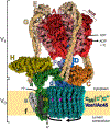
Fig. 1.
Fig. 1.
Structure and mechanism of the V-ATPase. The V-ATPase is organized into two large domains: the peripheral V1 domain, which hydrolyzes ATP, and the integral V0 domain, which transports protons. V1 is composed of subunits A, B, C, D, E, F, G and H. The A3B3 hexamer contains nucleotide binding sites at each of the six AB interfaces, with three of the sites performing ATP hydrolysis. Subunit A also contains a 100 amino acid non-homologous region (NHR) which is not present in the ATP synthase β subunit. The V0 domain is composed of subunits a, c, c′ (in yeast), c″, d, e, f, and Voa1 (homologous to mammalian Ac45). The complex operates by a rotary mechanism, with ATP hydrolysis causing rotation of the central stalk and proteolipid ring. Protons enter an aqueous hemichannel in subunit a and protonate glutamate residues (magenta color) in each proteolipid subunit. After performing a full rotation, protons exit through a second hemichannel upon stabilization of the glutamate side chain by a critical arginine (yellow Arg) in subunit a. The complex is always oriented with V1 in the cytoplasm, so proton transport is driven into lumenal compartments or the extracellular space. This 6.6Å cryo-EM structure of the Stv1p-containing, yeast V-ATPase holoenzyme was determined by Vasanthakumar et al. (PDB ID: 6O7V) [46].
Fig. 2.
Fig. 2.
Regulated assembly of the V-ATPase. An important form of V-ATPase regulation is the reversible dissociation of the V1 and V0 domains. In the disassembled state, free V1 cannot hydrolyze ATP, and free V0 cannot conduct protons. In free V1, HCT undergoes a large conformational shift, bringing it into contact with subunit F of the central stalk (PDB ID: 5D80) [45]. In free V0, the N-terminus of Vph1p or Stv1p (aNT) contacts subunit d of the central stalk (PDB ID: 6O7U) [46]. The only subunit that is released from either domain upon disassembly is subunit C. The process of reversible dissociation is rapid, does not require new protein synthesis, and occurs in response to diverse stimuli (see Table 1). In yeast, disassembly requires an intact microtubule network, while reassembly requires the RAVE complex, of which Rav1p is homologous to Rbcn-3A in higher eukaryotes. Other assembly factors common to yeast and higher eukaryotes include the glycolytic enzymes aldolase and phosphofructokinase (PFK). In yeast, PI(3,5)P2 binds to Vph1p and promotes assembly of Vph1p-containing V-ATPase complexes.
Fig. 3.
Fig. 3.
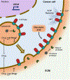
Function of V-ATPases in cancer cell survival, migration and invasion. Cancer cells upregulate a3 and a4, which target V-ATPases to the plasma membrane, where they transport protons extracellularly. An acidic extracellular pH causes protonation of cancer drugs (Drug+), which prevents their diffusion into the cell. Once in the cytosol, cancer drugs diffuse into acidic compartments, where they are retained upon protonation and prevented from reaching their intended targets. Plasma membrane V-ATPases also promote cancer cell survival by removing cytosolic acid produced from glycolysis. Plasma membrane V-ATPases may contribute to cell migration by creating regions of alkaline pH (↑pHi), which promote actin polymerization and branching near the plasma membrane. Alternatively, acidic extracellular pH may contribute to force generation at the leading edge through calcium influx. Finally, plasma membrane V-ATPases are thought to contribute to invasion by activating secreted proteases, which function to cleave extracellular matrix (ECM) components and to activate other secreted proteases such as matrix metalloproteases.
Fig. 4.
Fig. 4.
The V-ATPase participates in nutrient sensing at the lysosomal membrane. A) The V-ATPase associates with the Ragulator complex, which in turn is bound to the Rag GTPase heterodimer (Rags). When amino acids are abundant, Ragulator GEF activity towards the Rags enables recruitment of mTORC1 via its subunit Raptor (not shown). At the lysosome, mTORC1 associates with Rheb, which, when bound to GTP, stimulates the kinase activity of mTOR. V-ATPase inhibition or knockdown prevents mTORC1 lysosomal recruitment. V-ATPase assembly is also amino acid-dependent, with amino acid depletion leading to increased assembly, possibly as a way to increase protein degradation. mTORC1 activation by also requires growth factor (GF) signaling, which is independent of nutrient status. Growth factors bind receptor tyrosine kinases (RTK), leading to recruitment of PI3K, which phosphorylates PI(4,5)P2 (PIP2) to produce PI(3,4,5)P3 (PIP3). PIP3 recruits Akt to the plasma membrane where it is phosphorylated by 3-phosphoinositide-dependent protein kinase 1 (PDK1), enabling phosphorylation and full activation by mTOR complex II (mTORC2). Once active, Akt phosphorylates and inactivates the TSC complex, thereby relieving inhibition of Rheb. B) When glucose is abundant, the glycolytic enzyme aldolase (Aldo) is occupied by fructose 1,6-bisphosphate (FBP), and promotes Ragulator GEF activity towards the Rags. mTORC1 may then be activated by Rheb if growth factors are present (shown in A). Under these conditions, AMPK present at the lysosome is inactive, because its upstream kinase LKB1 is retained in the cytosol in complex with the scaffolding protein Axin. When glucose is low, Aldolase is no longer occupied by FBP, and Axin-LKB1 is recruited to the lysosome where it activates AMPK. Axin-LKB1 binding also appears to inhibit the Ragulator-Rag complex and displace mTORC1. The V-ATPase is required for glucose dependent recruitment of Axin-LKB1 to the lysosome. V-ATPase assembly is also glucose-dependent, with glucose depletion leading to increased assembly, possibly as a way to increase autophagic flux.

References

    1. Nishi T, Forgac M, The vacuolar (H+)-ATPases--nature’s most versatile proton pumps, Nat. Rev. Mol. Cell Biol. 3 (2002) 94–103. 10.1038/nrm729. - DOI - PubMed
    1. Forgac M, Vacuolar ATPases: rotary proton pumps in physiology and pathophysiology, Nat. Rev. Mol. Cell Biol. 8 (2007) 917–929. 10.1038/nrm2272. - DOI - PubMed
    1. Kane PM, The long physiological reach of the yeast vacuolar H+-ATPase, J. Bioenerg. Biomembr. 39 (2007) 415–421. 10.1007/s10863-007-9112-z. - DOI - PMC - PubMed
    1. Saroussi S, Nelson N, Vacuolar H(+)-ATPase-an enzyme for all seasons, Pflugers Arch. 457 (2009) 581–587. 10.1007/s00424-008-0458-9. - DOI - PubMed
    1. Breton S, Brown D, Regulation of luminal acidification by the V-ATPase, Physiol. Bethesda Md. 28 (2013) 318–329. 10.1152/physiol.00007.2013. - DOI - PMC - PubMed

Publication types

MeSH terms

Substances